Optimizing Patient Management with Ultra-Widefield Retinal Imaging
See More, Treat More Effectively
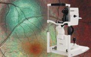
Ultra-widefield (UWF) optomap® imaging from Optos® is the first and only clinically validated non-contact technology able to image the peripheral retina (1). Combining SLO with patented ellipsoidal mirror technology, optomap acquires high-resolution images of both central and peripheral retina in one image across multiple imaging modalities, even in the presence of media opacities or pupils as small as 2 mm in diameter. But what impact has UWF optomap imaging had on clinicians’ practice and how they treat patients?
See more of the retina immediately
“UWF optomap imaging allows quick and easy examinations of the retina, which increases our understanding of the extent of our patients’ retinovascular and choroidal pathology,” explains Paulo Stanga of Manchester Royal Eye Hospital, Manchester, UK.
UWF optomap imaging gives you the ability to examine and assess nearly all of the retina (out to the ora serrata) in high resolution with its new montage tool. This is essential as many diseases, even those that were previously thought to affect the central pole only, manifest throughout the peripheral retina (2)(3). “The depth of field of UWF optomap imaging allows both the periphery and posterior pole to be in focus, and this is very valuable to us in documenting disease,” explains SriniVas Sadda of Doheny Eye Institute, Los Angeles, California, USA. “We’re recognizing that there are patients who have predominantly peripheral retinopathy with very little central disease, and these patients are at a substantially higher risk of progression due to proliferative disease” (2).
Clinical implications
UWF optomap imaging is useful not only for disease detection, but also for treatment planning and post-operative documentation. Several studies have indicated its utility in evaluating the success of treatment including placement of pan-retinal photocoagulation, sealing of holes, tears and detachments, and monitoring the impact of anti-VEGF therapy (4).
Avinash Gurbaxani of Moorfields Eye Hospital Dubai, UAE, and Antonia Joussen of Charité – Universitätsmedizin Berlin, Germany have both described the use of UWF optomap imaging to plan and monitor the success of peripheral laser treatment, while José García-Arumí of Instituto de Microcirugía Ocular, Barcelona, Spain considers UWF optomap imaging essential for surgical planning and post-operative monitoring in challenging retinal detachment cases.
Challenging cases
UWF optomap imaging, especially autofluorescence (AF), allows for the easy evaluation of otherwise challenging cases, including children and patients with rare inherited disorders such as familial exudative vitreoretinopathy (FEVR), retinitis pigmentosa (RP), and Coats’ disease.
Gurbaxani recalls a case where UWF optomap fundus AF imaging revealed RP in a two-year-old child with unexplained vision loss. “This child had been previously seen by several doctors who could not explain the cause of his poor vision as, clinically, his retina looked normal.” Gurbaxani finds UWF optomap imaging to be particularly useful when assessing the retinae of children, commenting, “It is easy for them to sit on the machine, it takes very little time and there is no bright flash – it has been invaluable in our clinic.” For Joussen, UWF optomap imaging has allowed her to more effectively evaluate FEVR, see peripheral vascular abnormalities associated with Coats’ disease, and identify and evaluate peripheral retinal tumors. She comments “This is where you need your Optos device to go to the periphery.”
The more you will see of the retina, the more you will diagnose and treat
UWF optomap imaging is becoming an essential part of many clinicians’ day-to-day practice, because the sooner ocular pathology can be seen, the earlier it can be treated. While the retina is fully visualized during clinical exam, having a static image of nearly the whole retina allows for zooming and manipulation of the image to allow for more effective assessment of small peripheral features that may have impact on treatment and management decisions. “The big picture view helps facilitate quick diagnosis – that is why UWF optomap imaging has become an indispensable tool in how we practice in our institution,” notes Sadda. Gurbaxani explains that “There are some pathologies we miss without UWF,” adding, “It has changed how I practice – I would not run a retina/uveitis clinic without it.”
Seeing more of the retina can provide greater insight and improve diagnosis and management. Stanga adds, “Without seeing, we cannot treat, so the more we see, the more we can treat. UWF optomap imaging has set the standard of care – it is difficult to imagine going back to standard fundus photography.”
UWF optomap imaging in clinical practice
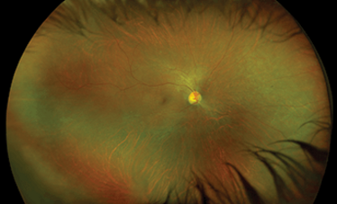
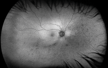
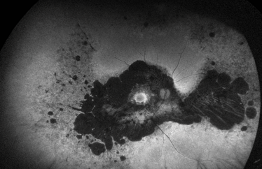
Retinal degeneration
Color and autofluorescence images of retinal degeneration, captured using the Optos California system
Courtesy of SriniVas Sadda, Doheny Eye Institute, Los Angeles, California, USA.
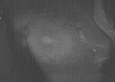
Retinitis pigmentosa
Autofluorescence image of retinitis pigmentosa captured using the Optos California system
Courtesy of David Brown, Retina Consultants of Houston, Texas, USA.
Pigmentary retinopathy
A 3-year-old child presented to Avinash Gurbaxani’s clinic with poor vision. The patient had received a prior diagnosis of Vogt Koyanagi Harada syndrome from a clinic in Spain and had been prescribed oral immunosuppression treatment. When referred to Gurbaxani for a second opinion, UWF optomap fundus autofluorescence imaging revealed a hyper/hypo autofluorescence pattern more consistent with inherited disease. Pigmentary retinopathy was later confirmed by genetic testing, saving the child from high-risk immunosuppression therapy.
Courtesy of Avinash Gurbaxani, Consultant Ophthalmic Surgeon in uveitis and medical retinal diseases at Moorfields Eye Hospital Dubai, UAE.
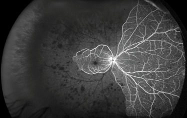
Ocular ischemia syndrome
Captured by UWF optomap fluorescein angiography imaging using the Optos California system
Courtesy of Paulo E. Stanga, Professor of Ophthalmology & Retinal Regeneration, University of Manchester Consultant Ophthalmologist & Vitreoretinal Surgeon, Manchester Royal Eye Hospital Director, Manchester Vision Regeneration (MVR) Lab at MREH and NIHR/Wellcome Trust Manchester CRF.

Color and autofluorescence images of
retinal degeneration, captured using the
Optos California system

Courtesy of SriniVas Sadda, Doheny Eye Institute, Los Angeles, California, USA.
- Optos (2016). Available at: bit.ly/optomap. Accessed August 30, 2016.
- PS Silva et al., Ophthalmol, 122, 949–956 (2015). PMID: 25704318.
- I Lengyel et al., Ophthalmol, 122, 1340–1347 (2015). PMID: 25870081.
- A Nagiel et al., Retina, 36, 660–678 (2016). PMID: 27014860.
