Subscribe to Newsletter
Business & Profession Refractive, Cornea / Ocular Surface, Imaging & Diagnostics

Iris Cysts

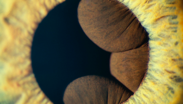
Credit: © Mark Maio www.markmaio.com

The image of confluent iris cysts was made on film using a photo slit lamp. The cysts were 360º and after making the proper medical documentation of the entire extent of the cysts, I moved the light source to the extreme left and positioned the light to fall tangentially across only half of the pathology to create a more visually pleasing image.

Receive content, products, events as well as relevant industry updates from The Ophthalmologist and its sponsors.

When you click “Subscribe” we will email you a link, which you must click to verify the email address above and activate your subscription. If you do not receive this email, please contact us at [email protected].
If you wish to unsubscribe, you can update your preferences at any point.

About the Author
Mark Maio

www.markmaio.com

Register to The Ophthalmologist

Register to access our FREE online portfolio, request the magazine in print and manage your preferences.

You will benefit from:
  • Unlimited access to ALL articles
  • News, interviews & opinions from leading industry experts
  • Receive print (and PDF) copies of The Ophthalmologist magazine

Register

Disclaimer

The Ophthalmologist website is intended solely for the eyes of healthcare professionals. Please confirm below: