Deep Learning Versus Keratitis
A new Lancet study highlights the accuracy of DL models for detecting and diagnosing corneal infections

Credit: AdobeStock.com
A systematic review and meta-analysis has explored the diagnostic capabilities of deep learning (DL) models for infectious keratitis (IK) – a leading global cause of corneal blindness.
Microbiological investigation is the current “gold standard” approach for diagnosing IK; however, in some countries – especially low- and middle-income countries (LMICs), which are disproportionately affected by IK – low microbiological culture yield, the need for clinical expertise in undertaking the investigations, and the long turnaround times associated with results emphasizes the need for alternatives.
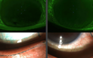
In the study, the multi-institutional team of researchers analyzed 35 studies, including over 136,000 slit-lamp/anterior segment photography (ASP) and in vivo confocal microscopy (IVCM) corneal images from more than 56,000 patients. The aim? Assessing the ability of DL models to identify and differentiate various forms of IK, including bacterial, fungal, viral, and Acanthamoeba keratitis.
Taken as a whole, the meta-analysis revealed that DL models demonstrated high diagnostic accuracy, with a sensitivity of 86.2 percent and a specificity of 96.3 percent in external validation studies. Furthermore, DL models were found to be as effective as ophthalmologists in diagnosing IK, with comparable sensitivity and specificity.
Another significant finding of the systematic review was that DL models, especially those based on IVCM images, showed excellent accuracy in distinguishing between different causes of IK, with a sensitivity of 91.8 percent and specificity of 94 percent. The use of IVCM allows for high-resolution imaging at a cellular level, making it especially useful for identifying challenging infections such as fungal or Acanthamoeba keratitis.
Though these results are promising, the study authors note that further investigations into data diversity are needed to fully realize the potential of DL in clinical settings. Additionally, the transparency and explainability of DL models must be enhanced to increase their acceptance among the clinicians who will be tasked with operating such tools.
The Ophthalmologist Presents:
Enjoying yourself? There's plenty more where that came from! Our weekly newsletter from The Ophthalmologist brings you the most popular stories as they unfold, chosen by our fantastic Editorial team!


