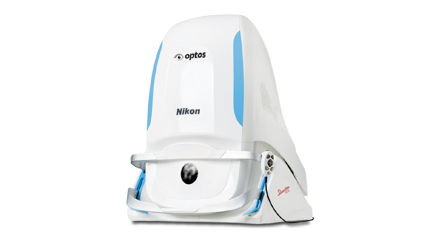
A core issue ophthalmologists routinely face in imaging is the ability to easily identify, localize, and structurally assess peripheral retinal lesions all within one single system. In a bid to remedy this daily workflow hurdle, UK-based optical device company, Optos, has developed the Silverstone RGB system. Launched in October 2025, the imaging system debuted at the American Academy of Ophthalmology (AAO) meeting in Orlando, Florida.
Rather than replacing the core architecture of Optos’ existing Silverstone platform, the upgraded RGB model retains Silverstone’s ultra-widefield (UWF) imaging plus lesion-guided swept-source OCT while also including nine imaging modalities. Combined, these modalities allow ophthalmologists to perform surface to deep tissue imaging (RGB, RG, red/green views), functional and metabolic imaging (FAF, BAF), vascular imaging (FA, indocyanine green angiography – ICG), and structural cross-sectional imaging (SS-OCT), all within the one system.
“Silverstone RGB was developed in response to a growing clinical need for more comprehensive, multimodal retinal assessment within a single, efficient platform,” explained Dana Keane, Vice President Clinical Affairs at Optos. “Traditional imaging approaches often require multiple devices or are limited in their ability to visualize the peripheral retina and deeper structures simultaneously. This integrated approach supports a more comprehensive assessment of pathology, more informed clinical decision-making, reliable longitudinal monitoring, and improved outcomes.”
Silverstone RGB differs from its predecessors, added Keane, in its combination of true-color, 200° single-shot ultra-widefield imaging with fully integrated swept-source OCT, which allows for "enhanced color fidelity, deeper tissue penetration, and the ability to evaluate pathology across every retinal layer, including the far periphery, with precision." Keane explained further: “Silverstone RGB’s integrated SS-OCT technology provides the ability to capture a 23mm line scan, as well as navigated volume scans, out to the edges of the UWF field of view. This enables clinicians to assess structure and pathology in a more complete and clinically intuitive way, improving both confidence and efficiency in diagnosis and management.”
Clinician feedback played a central role in the development of the new system, particularly in refining Optos’ understanding of how multimodal imaging could be most efficiently integrated into real-world clinical workflows.
Keane noted how, during the initial feedback rounds, eye care professionals consistently emphasized the need for three main components involved in this improvement: greater access to peripheral pathology without any added complexity, more intuitive ability to switch between different imaging modalities, and faster, high-quality image capturing that would create minimal burden on their patients.
“These insights informed the development of a platform that not only expands imaging capabilities, but also ensures those capabilities are accessible, efficient, and clinically meaningful in everyday practice,” she added.
