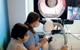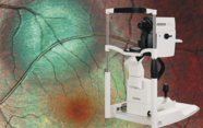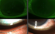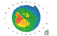
Over the last decade, intraoperative OCT has emerged as a valuable tool; not only can it provide real-time information on surgical outcomes, it can influence decision-making during the surgery. “Previous research has demonstrated the potential feasibility of intraoperative OCT when used externally to the microscope,” says Justis Ehlers of Cole Eye Institute of the Cleveland Clinic, Ohio, USA. Ehlers is Principal Investigator on the DISCOVER study, which was launched to evaluate the role of microscope-integrated OCT in ophthalmic surgery.
Building OCT directly into the microscope could bring several potential advantages over a separate system, such as increased efficiency and the ability to visualize tissue-instrument interactions. In this ongoing study, three prototype microscope-integrated OCT systems are being used by Cole Eye Institute surgeons to the feasibility and potential utility – the three-year outcomes of which have just been published (1). Of 837 eyes enrolled to date (244 anterior and 593 posterior segment cases), images were acquired successfully in 820 eyes (98.0 percent; 95% CI, 96.8–98.8 percent). In 106 anterior cases (43.4 percent; 95% CI, 37.1–49.9 percent) and 173 posterior cases (29.2 percent; 95% CI, 25.5–33.0 percent)surgeons reported that the technology influenced decisions during the surgical procedure. “We were surprised by the high frequency that OCT added value and impacted surgical decision-making – something that has also been confirmed in other studies,” says Ehlers.
According to the team, the three-year results demonstrate the feasibility and usefulness of microscope-integrated OCT. “I use intraoperative OCT for most of my surgeries, including macular cases, complex retinal detachments, and proliferative diabetic retinopathy,” says Ehlers. “I hope this study will help guide and inform surgeons about the potential impact of using the technology.” A multi-center randomized trial is apparently in the pipeline to provide critical information on comparative outcomes with intraoperative OCT. And Ehlers says that extensive work is continuing on enhancing the technology for image quality and tracking, as well as OCT-compatible instrumentation and software analysis platforms. Who knows what the surgeons of the future will be able to see as they operate.
- JP Ehlers et al., “The DISCOVER study 3-year results: feasibility and usefulness of microscope-integrated OCT during ophthalmic surgery”, [Epub ahead of print], (2018). PMID: 29409662.
