From differentiating geographic atrophy subsets, to determining the socioeconomic factors involved in retinoblastoma visual outcomes, these are the trailblazing studies that caught our attention this week…
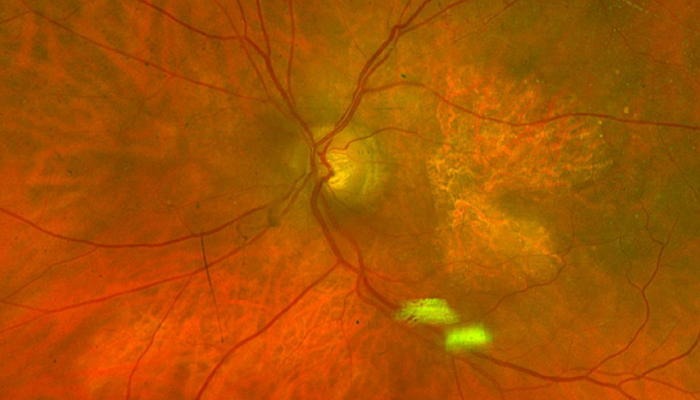
GA subtypes. Researchers behind an Ophthalmology Science paper have sought to determine the clinical similarities – and differences – between two forms of geographic atrophy (GA): conventional GA and pachychoroid GA. Examining patients from six Japanese university hospitals, the team identified a number of differences between the two subtypes, most notably that the progression rate of conventional GA was significantly higher than that of pachychoroid GA, and that pachychoroid GA tended to occur in a much younger, male demographic than conventional GA. Link
P-GAN positives. Artificial intelligence (AI) can increase the speed of retinal imaging by up to 100 times, according to the latest National Institutes of Health (NIH) research. Employing an AI method known as P-GAN (parallel discriminator generative adversarial network) to adaptive optics optical coherence tomography (AO-OCT), the research team was able to improve both retinal image contrast and the speed of diagnostic imaging times. They hope that this newly developed tool will help specialists better evaluate age-related macular degeneration (AMD), as well as other retinal diseases. Link
Retinoblastoma care inequities. To determine the links between socioeconomic factors and retinoblastoma visual outcomes, researchers from Mass Eye and Ear in Boston have conducted a retrospective study using data obtained from the IRIS Registry. Analyzing statistics of race, ethnicity, and insurance coverage, the team found poor visual outcomes were more likely in the Black and Hispanic populations, as well as in children who were publicly insured. Link
ICA risk factors. Investigating the association between diabetic retinopathy (DR) and the severity of internal carotid artery (ICA) stenosis, a BMC Ophthalmology study has suggested that the atherosclerosis caused by stenosis can contribute towards advancing proliferative diabetic retinopathy. These retinal changes – including retinal nerve fiber layer (RNFL) thinning – observed in ICA stenosis patients, the authors say, highlight the need for further research to be undertaken to determine the exact role of RNFL thickness in ICA stenosis patient management. Link

Credit: Fundus imaging of Geographic Atrophy on Retina by Ktkvtsh / CC BY
