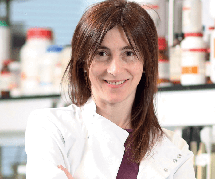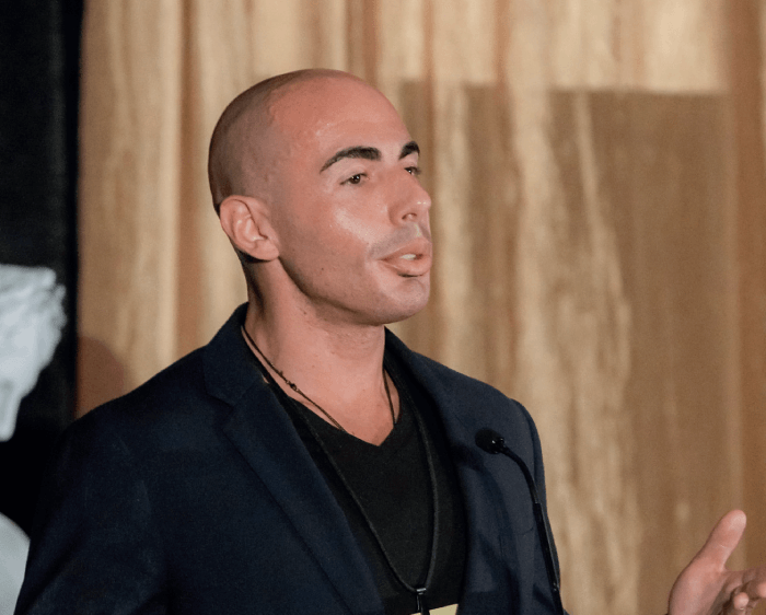
Noemi Lois is Professor of Ophthalmology, Queen’s University in Belfast, Honorary Consultant Ophthalmic and Vitreoretinal Surgeon, The Belfast Health and Social Care Trust, Belfast, Northern Ireland, UK
Route to the Retina: I was lucky to do subspecialty training in vitreoretinal surgery at the Royal Liverpool University Hospital in the UK, with David Wong. He is an outstanding and gifted surgeon: innovative, enthusiastic, very humble, and a fantastic teacher. I am very grateful to him for everything he has taught me.
Before that, I’d done an Ocular Oncology Fellowship at Wills Eye Hospital in Philadelphia (under the direction of Jerry and Carol Shields), and a Medical Retina Fellowship at Moorfields Eye Hospital in London (under the direction of Alan Bird). I was also very lucky to be able to do training in phaco-chopping techniques with an amazing cataract surgeon, Carlos Figueiredo, in São José do Rio Preto, Brazil. This experience helped me with phacovitrectomy – a very efficient surgical approach (when needed), which I very much enjoy doing.
David Almeida is a vitreoretinal eye surgeon and director of clinical research at Erie Retinal Surgery, a retina-only private practice in Erie, Pennsylvania, USA
Route to the Retina: My circuitous route sometimes feels like an adventure race spanning two continents and three countries! I completed medical school and ophthalmology residency at Queen’s University in Kingston, Ontario, Canada. Previously, I had finished a PhD in pharmaceutical chemistry at the University of Szeged in Hungary and an MBA in healthcare administration from The George Washington University School of Business. My aim was to blend my skill set and I was lucky enough to join the vitreoretinal service at the University of Iowa in Iowa City, Iowa for a two-year vitreoretinal surgery fellowship. Every day I am grateful for my journey to the retina and for the superb training and genuine friendships I developed along the way.

What do you consider to be the biggest milestones in surgical retina?
David Almeida: With respect to my career, generally, the introduction of transconjunctival sutureless vitrectomy (1, see the TSV box) accelerated efficiency gains with improved safety. Specifically, macular surgery, with the advent of Kelly and Wendel’s work on macular holes (2), provided opportunities for knowledge gains involving surgical approaches to membranectomy.
How have retinal surgery methods changed over the course of your career?
Noemi Lois: During my residency in ophthalmology, contact-viewing systems were widely used and vitrectomy was being done using 20-gauge incisions. When I started my vitreoretinal fellowship, wide-angle viewing systems were in place. These not only allowed for an excellent view of the fundus but also for “independence” – we do not need to rely any longer on an assistant maintaining and moving the contact lens for us, which is an advantage. New techniques were developed and their use became generalized, such as chromovitrectomy (see Chromovitrectomy box), which greatly facilitates surgical manoeuvers such as internal limiting membrane (ILM) peeling (see the ILM box). The use of ILM peeling was also generalized soon after I became a consultant vitreoretinal surgeon. It is incredible to think we are able to peel this extremely thin layer of the retina!
In recent years, small-bore vitrectomy was introduced. With this transconjunctival trocar system it is now hard to tell which eye has had a procedure even only a day or two following surgery, as there is minimal, if at all, chemosis and inflammation. Patients are very comfortable and the recovery is very fast. It has reduced our time to initiate and finalize the vitrectomy, allowing us to spend more time, if needed, inside the eye, doing the work.
David Almeida: I’ve enjoyed using valved trocar-cannulas (they provide excellent stability without the need for plugs) and lighted endolaser probes (allow for photocoagulation and scleral depression without an endoilluminator or assistant). Both of these solutions are simple, but they provide significant efficiency improvements; with a lighted endolaser probe, you can scleral depress for yourself and apply the laser at the same time without the need for an additional light source or assistant for scleral depression.
Transconjunctival sutureless vitrectomy surgery (TSV)
At the American Academy of Ophthalmology meeting in New Orleans, Louisiana, USA, in 2001 Gildo Y Fujii and colleagues presented a new 25-gauge instrument system for TSV (3). Based on the results of an experimental, comparative interventional in vitro study, they aimed to evaluate the infusion and aspiration rates and operative times of the surgery using 20- and 25-gauge vitrectomy system. They found that using 25-gauge TSV effectively reduced operative times that didn’t require full vitrectomy. In 2005, Claus Eckardt argued that 25-gauge instruments were too flexible for many of the complicated retina and vitreous body procedures, and proposed using a 23-gauge system (1).
What have been the biggest developments in this field over the last few years?
David Almeida: Over the last few years we’ve witnessed a myriad of refinements in vitrectomy for proliferative vitreoretinopathy and endophthalmitis. The challenges in these disease states require unique approaches, such as limbal vitrectomy (trocars placed in the corneal limbus) (5) and chromovitrectomy, which aid in addressing relevant pathology.
What techniques do you currently use?
Noemi Lois: I probably use most techniques that are currently around, although I like to see a good evidence base before introducing new techniques to my clinical practice, unless I use them within the context of a research study. The one technique I still do – and which many of us consider to be at “risk of extinction” – is scleral buckling (SB, see the Scleral Buckling box). I cannot think of anything else more rewarding than doing a scleral buckle for the appropriate retinal detachment. It is cheap and efficient: you can reattach the retina on the same day and, if that is the case, it is very unlikely the retina will detach later on. And the patient can go back to work right away, if needed. For the surgeon, it is fun to do, too.
David Almeida: I use the Alcon Constellation platform with either 25- or 27-gauge valved trocar-cannulas. I prefer the end-grasping ILM-type forceps which facilitate pinch-and-peel membrane removal of epimacular and peripheral membranes. I also use adjuvants like triamcinolone, methotrexate, and foscarnet for complex pathologies.
Internal limiting membrane (ILM) peeling (4)
Over the last couple of decades, ILM peeling has become a very common procedure. A combination of new microsurgical instruments and the availability of different dyes to stain the membrane have boosted the performance of ILM peeling, reducing both the time and trauma associated. ILM peeling has been used to treat various retinal pathologies, including full-thickness macular hole, epiretinal membrane, macular edema, vitreomacular traction syndrome, and Terson syndrome. ILM peeling is associated with better anatomical and visual outcomes after retinal surgery for the above conditions.
What are the biggest controversies or uncertainties of surgical retina?
Noemi Lois: There are still many uncertainties. However, our field continues to advance, and we have more evidence with regard to benefits and potential harms of our interventions than other fields of surgery. It is a complex area as there is a great deal of variability among people presenting with vitreoretinal disorders, which represents a challenge when designing randomized controlled trials. Despite this, we have managed to pursue them, and complete them successfully. For example, trials evaluating internal limiting membrane peeling and posturing for the treatment of idiopathic macular hole; trials comparing buckle or vitrectomy with pneumatic retinopexy and comparing buckle with vitrectomy for certain types of rhegmatogenous retinal detachment, just to mention a few. Nevertheless, there is still a great deal of work to do. I think it is essential that we put together our efforts through collaboration to ensure we will be able to address even the not so common problems (such as trauma, giant retinal tears, proliferative vitreoretinopathy).
David Almeida: Although not strictly a controversy, I worry about the decreasing use of scleral buckling – either as a primary technique or combined with vitrectomy – for retinal detachment repair. For some cases (such as young patients, high myopes, absence of posterior vitreous detachment), scleral buckling offers significant advantages. We actually published a “Scleral Buckling 101” article in The Ophthalmologist back in 2018 (6) to highlight case selection and pearls for successfully using the technique. I hope scleral buckling remains a fundamental tenet of vitreoretinal surgical training and remains relevant in the surgical literature.
Chromovitrectomy
In chromovitrectomy, the vitreoretina specialist uses dyes to better visualize diseases, such as macular holes and macular traction maculopathy, to achieve better postoperative outcomes. Blue and green dyes provide good contrast against the retinal pigment epithelium (RPE) natural color. Indications for chromovitrectomy include pars plana vitrectomy, ILM peek, ERM peel, macular hole repair, retinal detachment repair, and anterior vitrectomy. Dyes are normally injected intravitreally, either following removal of the intravitreal fluid through fluid-gas exchange, or straight into the intravitreal fluid (6).
What are the unmet needs in surgical retina – and the main barriers to addressing them?
Noemi Lois: Although challenges do remain when it comes to anatomically restoring the retina with surgery in various vitreoretinal conditions, a major challenge we face is to restore function and vision. For many years, I have concentrated on the repair of tractional retinal detachments and the removal of proliferative membranes in people with diabetes. I think this is one of the most challenging problems we face in vitreoretinal surgery today. Here, better intraoperative imaging technologies, including optical coherence tomography, would be very helpful; for example, to better discern planes for delamination. Robotic surgery may also allow improved control of surgical dissections and may provide input with regard to forces being applied to the retina, providing immediate feedback to the surgeon to avoid iatrogenic damage. However, in these cases, the successful removal of membranes and reattachment of the retina does not always equate to improved function and vision. Retinal ischaemia is a major event triggering the proliferation of diabetic membranes, which, through its growth and contraction, lead to tractional retinal detachment. Currently, there is no means to revascularize the retina. At the Wellcome-Wolfson Institute for Experimental Medicine in Northern Ireland, UK, I work with scientists to try to improve our understanding of retinal ischaemia and to develop therapies that may allow retinal blood vessel regeneration.
David Almeida: Implantation of retinal prosthetic devices, pioneered by Humayun and colleagues (11), represents an important advancement in retinal surgery. However, this is limited to a few sites, given the surgical learning curve and technical requirements. I expect there will be additional retina prosthetic devices, along with stem cell products, approved in the future, and these will require further refinements to how vitreoretinal specialists approach surgery.
Have you applied robotics in your practice?
David Almeida: A fascinating question. I do not currently use robotics as a means of executing procedures, but this may change. I foresee a major limitation being the three-dimensional spherical nature of the eye and the unique challenges of working in spaces such as the anterior chamber or posterior segment. I am interested in the concept of robotics employed as surgical assistants, and I envision vitrectomy platforms with robotic components to directly aid retina surgeons with respect to instruments, tamponade agents, and similar aspects of retinal procedures.
Scleral buckling (7)
Scleral buckling (SB) was first described in 1949 by Ernst Custodis, and was further popularized by Charles Schepens and Harvey Lincoff in the 1950s. Over the past six decades, SB principles and techniques have remained relatively unchanged. The technique favorably alters the geometry and physiology of the eye to help close and maintain closure of retinal breaks. Inward indentation of the eye in conjunction with externally applied cryotherapy or laser photocoagulation creates a permanent adhesion between the neurosensory retina and the RPE. Furthermore, SB-induced indentation helps overcome the forces tending to detach the retina, including cellular epiretinal proliferation and the magnitude and direction of vitreous traction on the neurosensory retina (8). SB surgery is advantageous because, as well as treating existing retinal breaks, it also supports the vitreous base, which prevents new retinal tears (9). Additional advantages of SB over PPV include a lower incidence of cataract (which may help preserve accommodation in younger patients), fewer complications (such as endophthalmitis or choroidal hemorrhage), and no need for post-operative positioning or travel restrictions (10). But in this age of vitrectomy, which patients are best candidates for a primary scleral buckle?
Scleral buckling surgery should be strongly considered in patients presenting with specific scenarios, which are outlined below with our reasoning:
- Young, phakic patients with no posterior vitreous detachment. Why? Avoids cataract formation. Moreover, induction of a posterior vitreous detachment (PVD) during PPV can be technically challenging and create iatrogenic retinal breaks.
- Retinal dialysis. Why? Typically, there is no associated PVD with dialysis. Further, given its anterior location, it can be difficult to visualize and therefore perform adequate vitrectomy in the area of dialysis.
- Very anterior break(s). Again, it is challenging to treat anterior retina with PPV.
- Patients with extensive lattice or multiple retinal breaks at the vitreous base. SB provides 360° support to the vitreous base and peripheral retina thereby preventing future tears.
- High myopia with contact lens intolerance in phakic, middle-aged patients with minimal or no cataract. SB will not cause significant cataract acceleration or anisometropia; however, PPV will accelerate the formation of cataract, which could complicate cataract surgical planning, as the most attractive refractive outcomes will induce significant anisometropia.

How about your biggest frustrations as a retina surgeon?
Noemi Lois: As for any surgeon, I guess it is not being able to help all patients. Not being able to always successfully reattach a retina with a single surgery. Not being able to understand why, for example, despite doing a good vitrectomy and closing all visible retinal breaks or holes, a retina still re-detaches. Frustration is unhelpful, however, so we should ensure we turn “frustration” into “challenge.” A challenge is stimulating and will lead us to success.
David Almeida: To avoid a downward spiral, I will say “valuation” of vitreoretinal surgery is a major frustration I have. How retinal surgery is evaluated, remunerated and coded is incongruent with the skill and intensity required. And that creates a disincentive for retina specialists to perform surgery and hinders innovation in new techniques and products.
What exciting projects, research or developments in surgical retina are you following? How are they likely to change the field?
David Almeida: I’m interested in the use of adjuvants to aid vitrectomy for complex pathologies; for example, triamcinolone chromovitrectomy for retinal detachment repair (12), foscarnet for complex retinal detachments secondary to viral retinitis (13), and fibrin glue for optic disc pit maculopathy (14). I believe adjuvants like foscarnet or methotrexate have the ability to significantly mitigate complex pathology that surgery alone may be insufficient for. We are currently working on methotrexate for proliferative vitreoretinopathy.
What do you see for the field’s the future?
David Almeida: In the short term, I believe we will see further improvements in enhanced surgical visualization along the lines of the Ngenuity 3D Visualization system (Alcon) and intraoperative optical coherence tomography imaging. Moreover, surgically implanted delivery devices for vascular endothelial growth factor inhibitors and surgical approaches for stem cell delivery will provide new insights in how we can modulate the eye to affect different disease states. In the longer term, efforts will continue to be directed at improving the efficiency of retina surgery with respect to operating room design, technology aids, and logistical integration.
What is your ultimate wish list for the field of surgical retina?
Noemi Lois: The ability to undertake “regenerative” surgery. As I mentioned above, being able to regenerate retinal blood vessels would transform outcomes for people with proliferative vascular disorders, including proliferative diabetic retinopathy. The ability to transplant retinal cells, of any kind, with the goal of regenerating damaged or lost ones is exciting. Induced pluripotent stem cell technology makes this a real possibility, bypassing problems related to the harvesting of cells and rejection. However, whatever we do – and whichever surgical technique we develop – we need to ensure they are easy enough for any trained vitreoretinal surgeon to perform, safe and efficacious and, very importantly, acceptable to patients. Ticking those three boxes would guarantee their implementation in clinical practice and their accessibility to patients.
David Almeida: I have two! First, preservation of robust training programs to ensure ongoing innovation in vitreoretinal surgical techniques and retainment of key procedure skills. Second, productive collaboration on patient needs between surgeons and industry is fundamental to facilitating new surgical approaches (such as stem cell delivery), which I am hoping will flourish.
If you could go back and give yourself one piece of advice at the start of your career, what would it be?
Noemi Lois: The one I gave myself so many years ago: “Do not worry about spending time training and working hard to pursue the best possible learning experience. It will be worth it and the benefits will be long lasting.”
David Almeida: “Never use two steps when one will do!” Economical surgical manoeuvers will not only improve your efficiency in the operating room, it benefits patient safety by minimizing surgical time. Additionally, a focus on eliminating surgical redundancy provides vantage of “pain points” inherent in current surgical approaches and techniques – and that aids innovation (15).

References
- C Eckardt, “Transconjunctival sutureless 23-gauge vitrectomy,” Retina, 25, 208 (2005). PMID: 15689813.
- NE Kelly and RT Wendel, “Vitreous surgery for idiopathic macular holes: results of a pilot-study,” Arch Ophthalmol, 109, 654 (1991). PMID: 2025167.
- GY Fujii et al., “A new 25-gauge instrument system for transconjunctival sutureless vitrectomy surgery,” Ophthalmology, 109, 1807 (2002). PMID: 12359598.
- E Abdelkader, N Lois, “Internal limiting membrane peeling in vitreo-retinal surgery,” Surv Ophthalmol, 53, 368 (2008). PMID: 18572054.
- K Xu, EK Chin, DRP Almeida, “Five-port combined limbal and pars plana vitrectomy for infectious endophtalmitis,” Case Rep Ophthalmol, 7, 289 (2016). PMID: 28101048.
- AAO, “Chromovitrectomy” (2020). Available at: https://bit.ly/2HiYisM.
- A Ringeisen, D Almeida, “Scleral Buckling 101,” The Ophthalmologist (2018). Available at: https://bit.ly/2FGxq5L.
- D Bloch et al., “The mechanism of the cryosurgical adhesion. III. Statistical analysis,” Am J Ophthalmol, 71, 666 (1971). PMID: 5546312.
- DJ D’Amico, “Clinical practice. Primary retinal detachment,” N Engl J Med, 359, 2346 (2008). PMID: 19038880.
- JB Conart et al., “Results of scleral buckling for rhegmatogenous retinal detachment in phakic eyes,” J Fr Ophtalmol, 36, 255 (2013). PMID: 22981521.
- MS Humayun et al., “Interim results from the international trial of Second Sight’s visual prosthesis,” Ophthalmology, 119, 779 (2012). PMID: 22244176.
- DRP Almeida et al., “Multiplane peripheral vitreous dissection with perfluoro-n-octane and triamcinolone acetonide.,” Retina, 35, 827 (2015). PMID: 25768254.
- K Xu et al., “Intravitreal foscarnet with concurrent silicone oil tamponade for rhegmatogenous retinal detachment secondary to viral retinitis,” Retina, 36, 2236 (2016). PMID: 27429388.
- DRP Almeida DRP, et al., “Fibrin glue and internal limiting membrane abrasion for optic disc pit maculopathy.,” Ophthalmic Surg Lasers Imaging Retina, 49, e271 (2018). PMID: 30566713.
- DRP Almeida, Retina Specialist, “Never use two steps when one will do,” (2019). Available at: https://bit.ly/3o7FEFd.
