Visual performance is often distilled from the health of the eye – with acuity changes occurring as a direct result of eye conditions, such as amblyopia. But what if you could improve vision without targeting the physiology or anatomy of the eye? My team and I have further developed technology to do just that. By using a web-app based program that trains the visual portion of the brain, we’ve proven that vision can be improved by an average of 2.5 lines on the visual acuity (VA) scale and contrast sensitivity function (CSF) can improve by 100 percent (post-cataract surgery patients can return to the normal range of CSF – a 150 percent increase). These improvements can be gained at home through a course of 30–40 half-hour sessions over a two to three month period.
In short, our program enhances vision by probing specific neuronal interactions, using a set of patient-specific stimuli that improve neuronal efficiency and induce improvement of Contrast Sensitivity Function (CSF), due to a reduction of surrounding neural noise and an increase in signal detection point. The improved contrast sensitivity results an improvement in visual acuity, and also stereo acuity in amblyopia.
Standing on the shoulders of giants
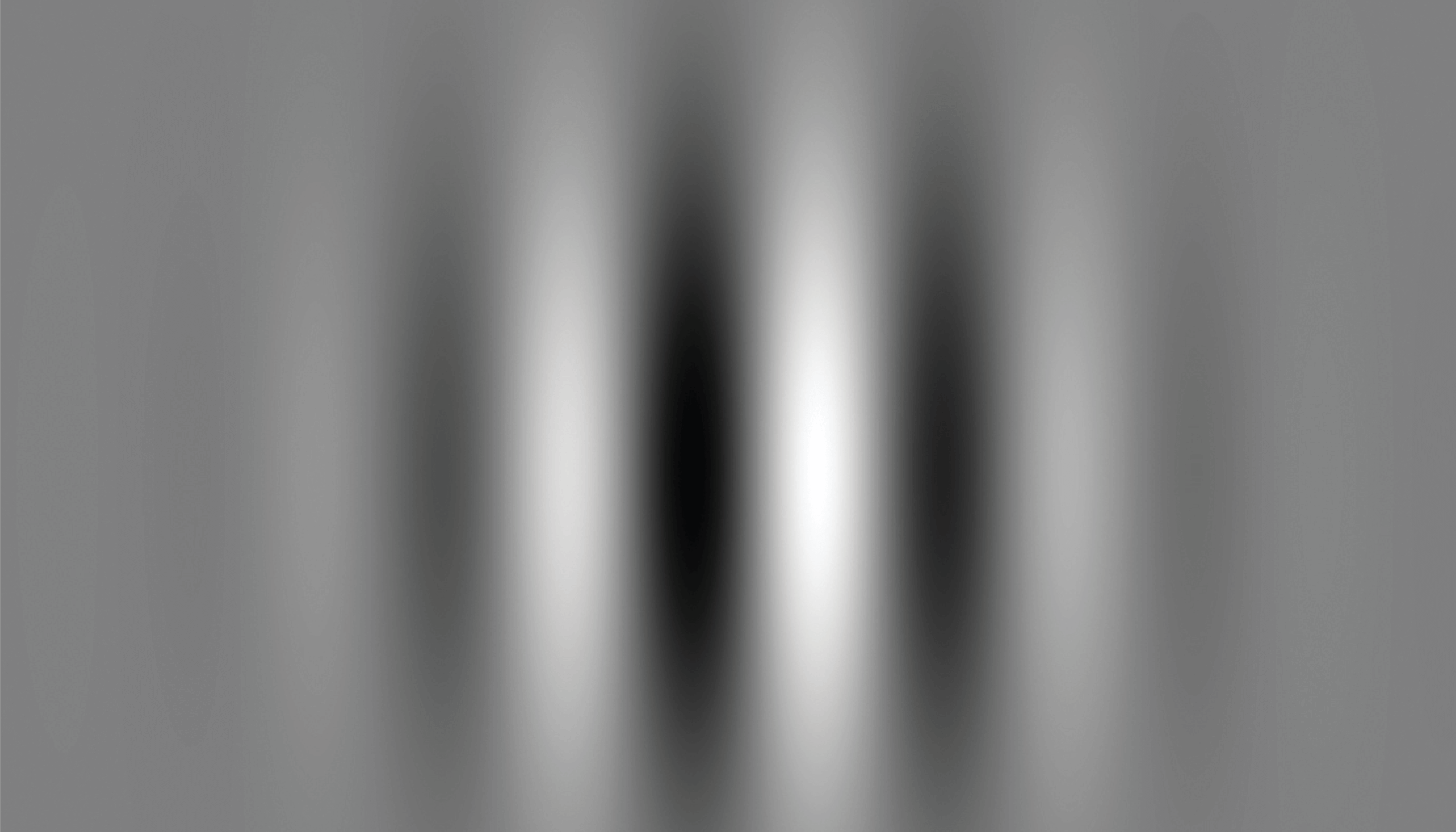
The scientific principles at play stem from the work of several Nobel Prize winning scientists. First up is Dennis Gabor, who created the hologram and, importantly for us, the Gabor patch (see Figure 1). The patch image has a mathematical formulation and shape that perfectly matches the receptive field of neurons in the visual cortex – and it induces a high level of stimulation for this area (2). Second, we have David Hubel and Torsten Wiesel’s prize-winning discovery that individual neurons respond to visual stimuli of precise location, orientation, and spatial frequency – and also that collective neuronal interactions are responsible for image characterization (3). And that basically means that we can use specific images to target and stimulate specific individual and collective neurons to improve vision for the patient. The final aspect of scientific background to the technology comes from discoveries made by Uri Polat: who demonstrated how neural respond of specific neurons in the primary visual cortex can be effectively increased through Lateral Masking Technique - placing Gabor patches at specific flanking positions, which masks the central Gabor target. The response to stimuli presented within the receptive field can be facilitated or suppressed by other stimuli falling outside the receptive field which, when presented in isolation, fail to activate the cell (4). In a later controlled randomized study, published in PNAS (5) Uri Polat demonstrated how this technique improves vision in adults with amblyopia beyond the critical period with lasting effect.
All of this knowledge allows us to take advantage of the brain’s amazing plasticity – to both diagnose and treat visual issues. Using this Lateral Masking Technique with Gabor patches of a different location spatial arrangement, contrast, orientation, frequency, and exposure duration we can stimulate and train individual and collective neurons to improve vision tailored specifically to the patient’s own visual deficits. These discoveries led to the establishment of a company in 1999 that used this technology to treat amblyopia – and has more recently led to the revamping of the technology, into a platform that is friendly to patients and clinicians, and the company as we know it today.


Plastic fantastic
In fact, almost all humans can improve their visual performance by capitalizing on the remnant's neuroplasticity that exist in adults . But those with visual impairments clearly stand more to gain. For example, amblyopia was previously only treatable at a critical period in youth (under nine years of age); our technology is unique in its ability to improve vision in adulthood – and there’s perhaps no greater proof than FDA approval to treat amblyopia in patients over the age of nine, with no upper age limit!. Recently, the American Medical Association has approved 2 new unique CPT codes for amblyopia treatment, using RevitalVision software.
Although the technology is designed to treat one condition, by applying it to people without a cortical defect (for example, those with a minor refractive error or physical damage to the eye) we still see improved processing of images in the brain, which compensates for the blurry images being transferred from the retina. In essence, any patient with a low-vision condition can improve their eyesight through visual cortex training.
How does the technology work?
With a digital camera, the quality of the final image depends on two main factors: image capturing and image processing. Therefore, if you use a digital camera with an imperfect lens but improve the processor, you can access an image with better resolution. Similarly, the quality of the image seen by humans is dependent on how the image is transferred from the retina to the brain and subsequent visual processing. If you can’t improve the anatomy of the eye any further, vision can still be improved by working on the processor.
Another analogy is fitness training – but instead of the effects of your hard work wearing off when life gets in the way and you can’t train for a while, the neural connections built over the sessions stay. In that way, I suppose it is more comparable with learning to swim or ride a bike – the visual improvement is retained for years when it is improved through the brain.
Life changing improvements
The exciting thing about this technology is the broad range of people it can help. For example, consider a patient with congenital nystagmus as a consequence of albinism, who hasn’t been able to acquire a driver’s license; here, a couple of lines gained in VA could be life changing. We have seen people who are amazed at being able to see the subtitles on TV at home for the first time, which allows them to sit at a normal distance away from the screen. I remember an 83-year-old patient with one remaining eye, who had vision loss when she was young and had suffered from dry AMD for the last 20 years; she went from 20/100 to 20/40 vision. We have seen candidates for air force pilot course who pass all their examinations – apart from the vision exam – gain the boost needed to qualify. And we have also seen patients with Stargardt disease who retained their improved VA for at least 12 months.
When you look at the original target for treatment: people with amblyopia, which has no alternative treatments, you can really see the benefit.
It’s a marathon, not a sprint
In developing the technology, we’ve faced a few obstacles – not least the need to start from scratch to transform it from an old windows based technology to a modern web-app to make the program and technology platform user friendly. We thought the project would take around six months – three years later, we were getting close to completion. In particular, the algorithm itself proved to be a major hurdle; the clinical and regulatory claims were all based on those original algorithm – so we had to reconstruct it perfectly. Another burden was financial – why should investors pay into a technology that failed to reach the market after investment of $35 million by the previous owners of the company. Thankfully, by explaining the exact problem and how we were going to overcome it, we were able to gain investors. Due to the company being focused on research and development, and the rehaul that was needed on the old platform to make the treatment user friendly for clinicians and patients, our work has somewhat flown under the radar. Whereas the old platform was used because it was the only option, now that the new technology platform is finished, we're ready to hit the ground running.
Now, the main challenge ahead is the ongoing education of the market – especially when the treatable visual system is more or less perceived to end at the retina or the optic nerve. We will continue to highlight the value of our technology to all potential “customers” – which includes eye care professionals, patients, and payers in the US. Despite the brain being seen as a black hole (and despite neuro-ophthalmology often being more bench than bench-to-bedside), we believe the visual cortex and visual processing should be brought into modern eye care for one simply fact: it’s proven ability to improve vision in line with patient needs.
In our mission, we are trying to reach as many people as possible all over the world. We currently have customers in North America, Australia, China, Korea, India, and the Middle East –and we also have clinics that work with us directly all over Europe. Given our focus on research and development over the past three years, we have been working somewhat under the radar – but now we are very much looking forward to launching our system in the US and working with regulators in other markets.
Seeing is believing
We have a proven way to benefit from the plasticity of the human brain – in a relatively short time frame. And I can see no reason to neglect treating the brain when vision is involved; we have clinically proven that visual performance is not eye deep. I firmly believe that our treatment will become a standard part of care for treating other diseases and visual conditions. To that end, we want to expand the FDA approval to other conditions (currently, it only covers amblyopia for ages nine and above).
If you’re reading this and in any way dubious about the technology, I am very keen to point you to our solid base of research and evidence. We find that skeptical ophthalmologists typically change their stance after a short presentation outlining the scientific principles and clinical data, but they often want to check it out for themselves – and we encourage all good doctors to do the same! After all, “seeing is believing.”
References
- D Gabor, “Theory of Communication,” Journal of the Institute of Electrical Engineers, 93, 429 (1946).
- JG Daugman, “Two-dimensional spectral analysis of cortical receptive field profiles,” Vision Res, 20, 847 (1980). PMID: 7467139.
- DH Hubel, TN Wiesel, “Receptive fields of single neurons in the cat’s striate cortex,”. J Physiol, 148, 574 (1959). PMID: 14403679.
- U Polat et al., “Collinear stimuli regulate visual responses depending on Cells contrast threshold,” Nature, 391, 580 (1998). PMID: 9468134.
- U Polat et al., “Improving vision in adult amblyopia by perceptual learning,” PNAS, 101, 6692 (2004). PMID: 15096608.
