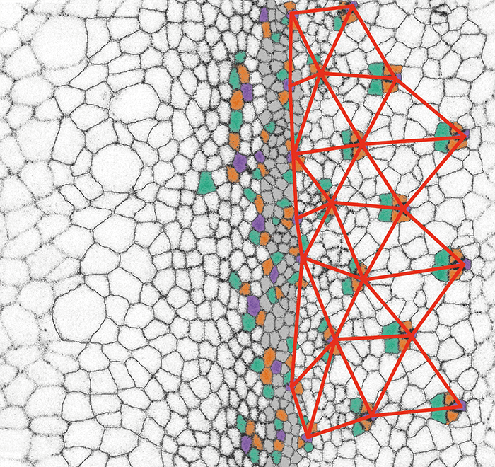
 Visualization of the mechanical wave that starts at the gray epithelium during the development of the fruit fly eye hexagonal pattern (marked in red). Credit: Northwestern University
All biological structures have an amazing process of development over time, eventually becoming a functional tissue, or – in some cases – a dysfunctional tissue! The structure of each tissue is crucial to function – whether that be the drainage allowed by the shape of the trabecular meshwork, the wings of a butterfly, or the processes of our spinal cord. The eye is not exempt from the wonders of developmental biology, and researchers from Northwestern University have tracked and investigated the stimuli that enable the eye to form as a functional tissue.
Using live imaging, the development of a fruit fly eye was tracked, with a dynamic movement of cells – opposing what was previously believed to be a relatively static process. The process uses both chemical signals between cells and mechanical forces to form the final tissue structure. This mechanical “wave” from the retina is marked in red within the featured image, and shows how the distinctive hexagonal form of the fruit fly eye develops from the gray epithelium at the center.
The researchers used time-lapse microscopy to get the amazing images – see the video below. Cells move in a flowing profile to the anterior, where they alternate in groups within each wave to slow down and leave at the edge of the wavefront. Molecular biologist Richard Carthew of Weinberg College of Arts and Sciences commented, “There is a constellation of patterns everywhere you look.” This pattern can also be compared to the directional engineering of crystals in engineering contexts.
Customized live-imaging tools enabled the tracking of eye development, and a computational tool developed in groups allowed identification and tracking of singular cells. Credit: Northwestern University
Are there any aspects of human or animal biology that you find fascinating? Or do you have any remarkable images of the majesty of tissue development and structure? We’d like to know or see them! Contact us at edit@theophthalmologist.com.
References
- KD Gallagher, M Mani, RW Carthew, “Emergence of a geometric pattern of cell fates from tissue-scale mechanics in the Drosophila eye,” Elife, e72806 (2022). PMID: 35037852.
