Until recently, the mechanisms that regulate the number of cells in the lens and, accordingly, its size and shape, have remained a mystery. Now, a group from Washington University School of Medicine has published the results of nearly four years of research into growth regulation in mouse eyes. Steven Bassnett and his laboratory examined the dynamic relationship between cell proliferation in the epithelial layer and macroscopic growth of the lens. To do this, they used confocal microscopy to track the locations on the lens surface where cells were in S phase – the point in the cell cycle at which DNA is replicated to prepare for cell division.
The researchers discovered that mitosis was largely restricted to a narrow segment of equatorial epithelium known as the germinative zone (1). As new cells are formed in the germinative zone, they pushed previously formed cells toward the equator of the lens; cells at the equator were pushed away from the lens’ surface and into its center. The number of cells in the lens epithelium continued to increase until the mice were about four weeks of age, peaking at 50,000 – but even after that number began to decline, the lens continued to grow. This, the researchers concluded, was due to a continued increase in the surface area of all cells, whether newly replicated or older.
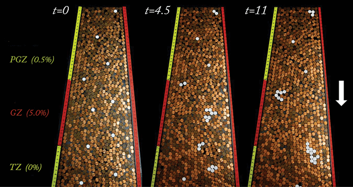
Having determined the way cells replicate in the lens, the Washington University group realized that it was similar in nature to the movement of coins in the Penny Pusher carnival game (Figure 1). In Penny Pusher, players add coins to a moving platform already covered in older coins, pushing the oldest coins off the platform’s edge onto a lower, larger platform and, ultimately, into a collection area where the player can access them. “We made a physical model of the lens equator using layers of pennies to simulate the division and migration of the lens cells,” Bassnett told the Association for Research in Vision and Ophthalmology. “Our Penny Pusher model looked very similar to [the carnival game].” The model was simplified, considering all cells to be of a single type whose proliferative behavior depended solely on latitude. Despite this, though, it was able to predict the emergence of cell clones, and its simulations were in good agreement with data obtained from lineage-tracing studies in mice.
The next step for Bassnett’s group is to examine the effects that these cells could have on the lens as a whole. With relatively few cells responsible for replication, mutations could have a multiplicative effect. “We are currently examining whether mutations in the DNA of individual lens cells can be transmitted to large numbers of lens cells, potentially influencing the clarity of the tissue and resulting in cataract,” explained Bassnett. Whether exploring the size and shape of the lens or analyzing the effects of proliferative cell mutations, it seems that the simple, stochastic Penny Pusher model offers valuable new insights into lens growth and regulation.
References
- Y Shi et al., “The penny pusher: a cellular model of lens growth”, Invest Ophthalmol Vis Sci, 56, 799–809 (2014). PMID: 25515574.
