Eyes are invariably considered objects of beauty, as well as the main instruments through which to perceive it. The scientific world has also been fascinated by these extremely complex organs, allowing our understanding to evolve greatly over time – a process you can observe in Ophthalmology Through the Ages below. Today, supported by advanced imaging technology, we have more ways of investigating eyes than ever before – and yet they remain mysterious and awe inspiring. If you are here to explore some of the many ways artists, researchers, and clinicians view eyes, you’re in for a real treat.
Mark Erickson has been a self-taught ophthalmic illustrator and animator since 1998. All of his art is inspired by his experiences working as an ophthalmic photographer since 1988. He says: “I’ve been lucky enough to observe, photograph and film the eye's anatomy, diseases and conditions, and surgery, in just about every possible angle or medium! When I’m at a slit lamp or photographing an eye, I can’t help but see the subject as a canvas with boundless artistic potential.”
Erickson’s artwork starts with a paper pencil sketch, which is then scanned into a digital painting tablet where he creates all his eye illustrations and digital paintings.His work has appeared on more than 50 ophthalmology journal covers and National Geographic books. He has been inspired by the realist style of Andrew Wyeth and the great impressionist, John Singer Sargent.
You can see more of his artwork at http://eyeimages.etsy.com and www.JirehDesign.com
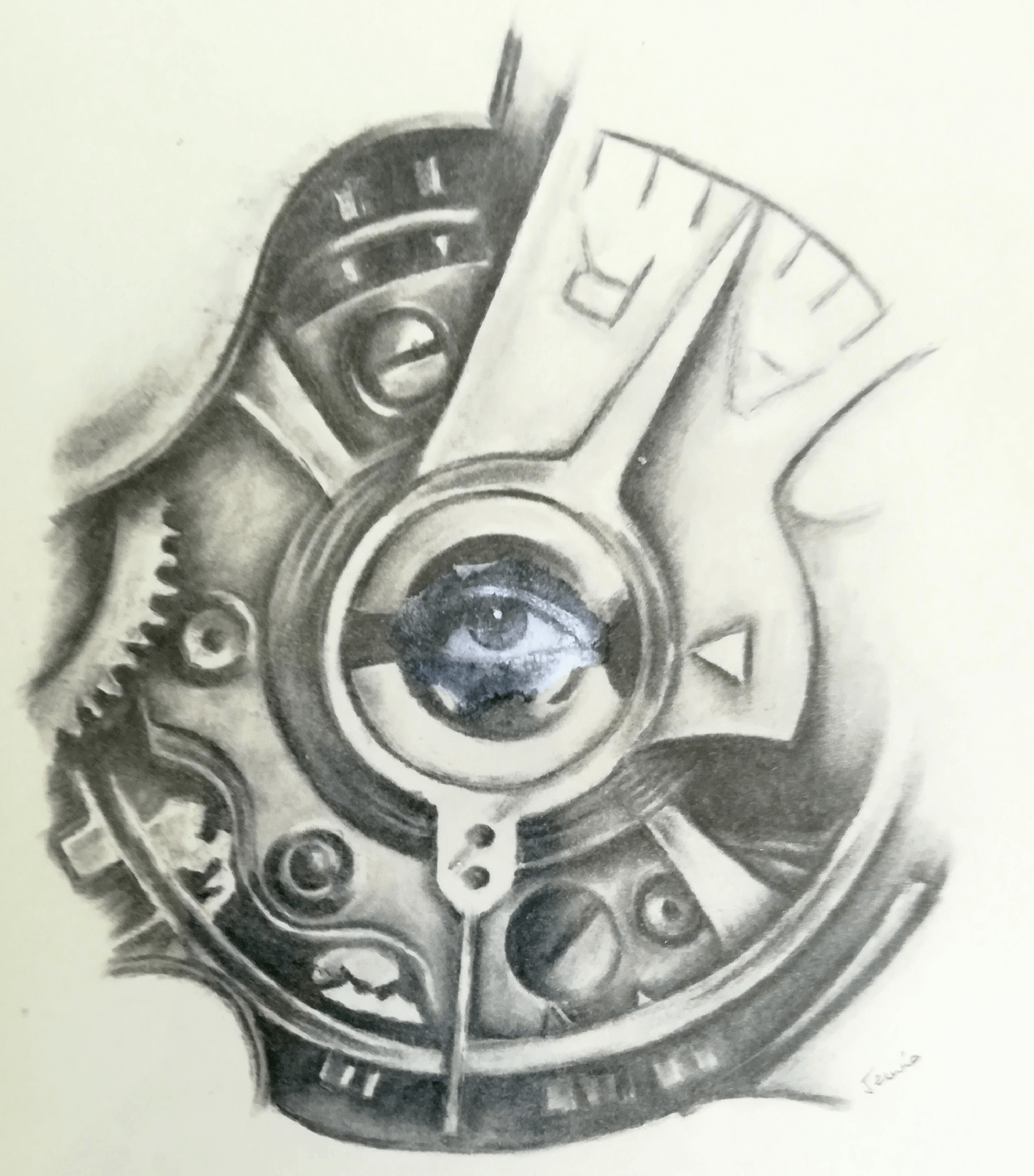
Peter Maloca is a double Wellcome Image Award winner for the best in science image making across the globe. He works as a medical doctor, eye surgeon and is Associate Professor at the University of Basel, Switzerland. He is also Group Leader Ophthalmic Imaging at the Institute of Molecular and Clinical Ophthalmology in Basel and works with Moorfields Eye Hospital in London, UK.
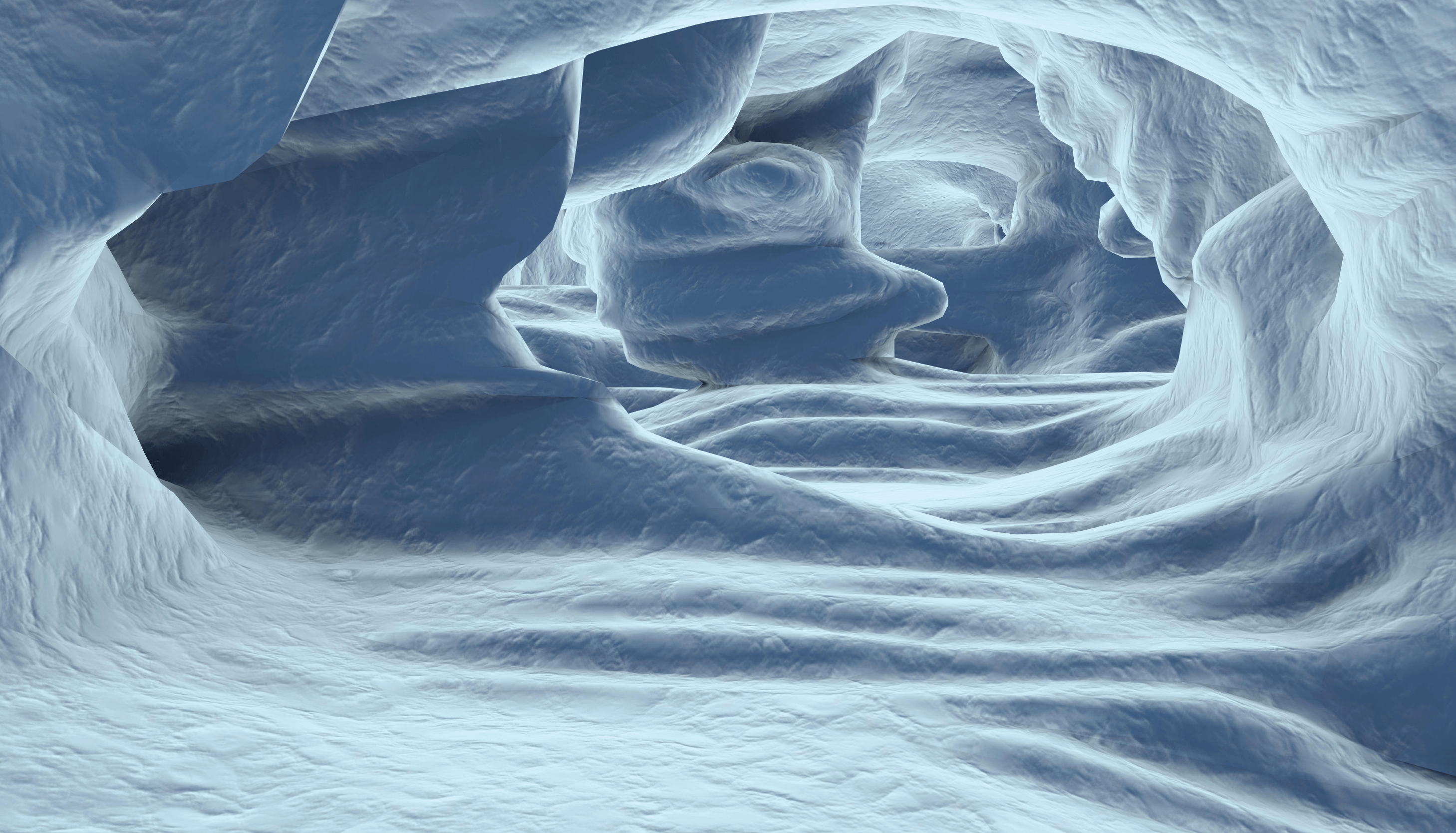
His special focus is medical retina and its fantastic 3D visual displays in modalities such as virtual reality. He says: “Nothing is more touching than the inner universe of the eye.”
Jui Telavane is a final year ophthalmology resident at Pravara Institute of Medical Sciences in Ahmednagar, India.
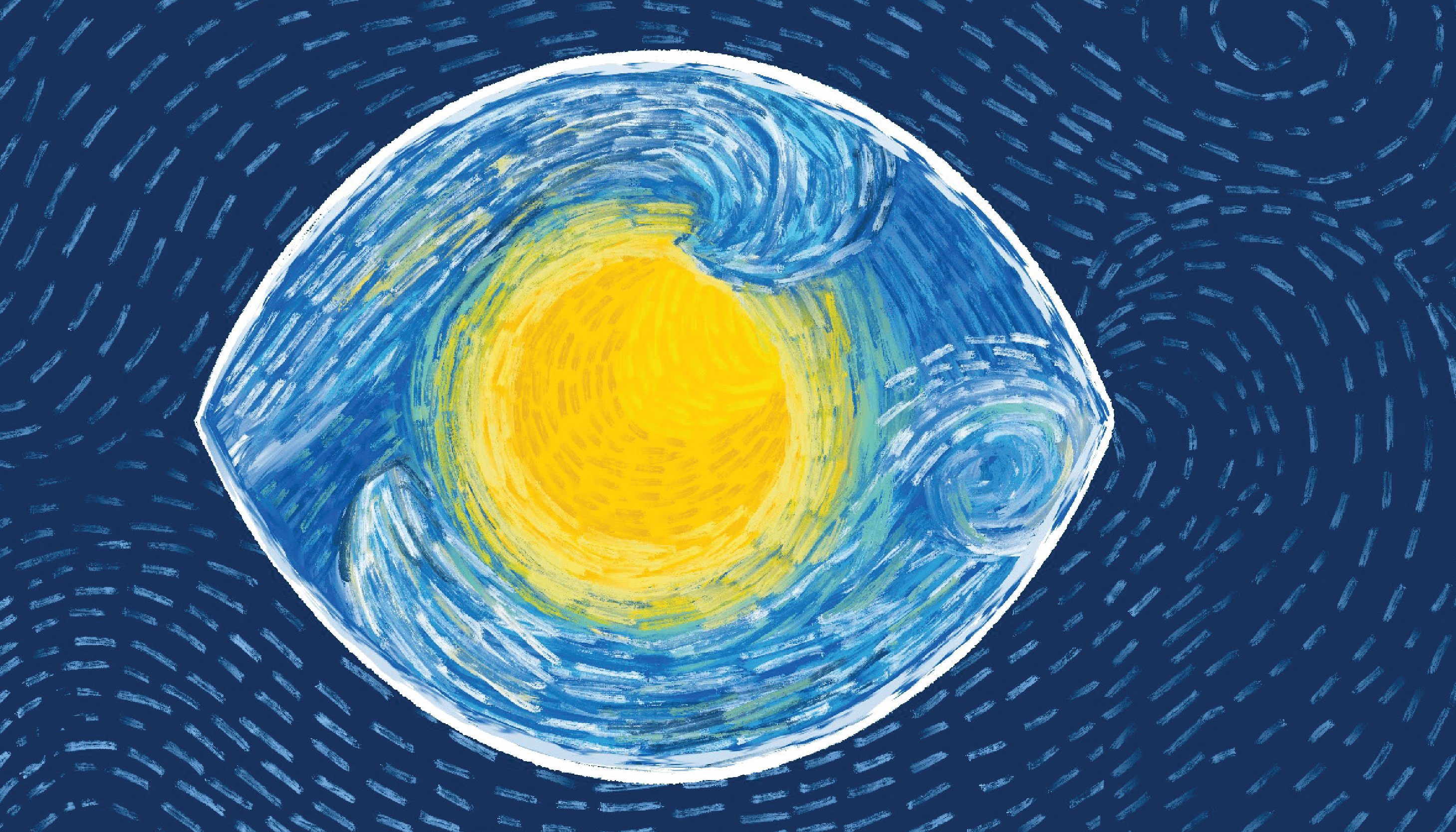
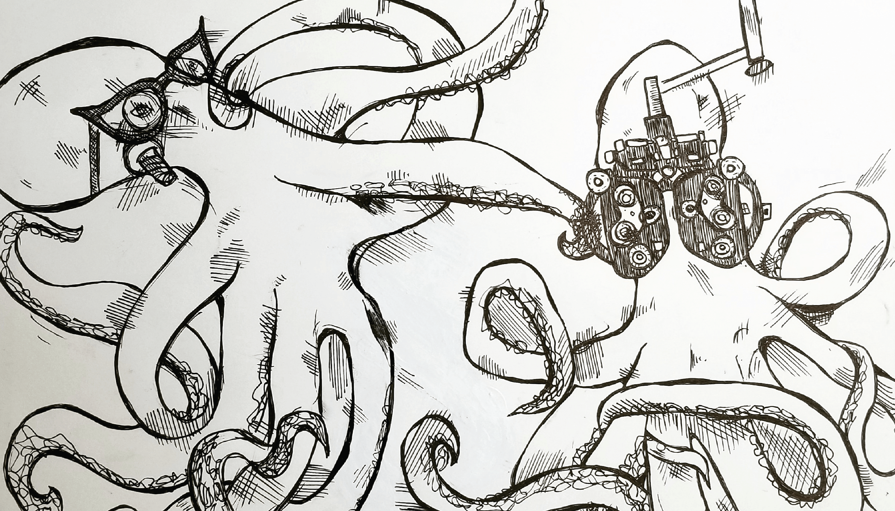
She comments: “I have always had a wacky imagination and I enjoyed art and drawing until I entered medical school, got busy, and lost touch with it. The pandemic slowed down life and drew me back to doodling. I was able to appreciate and connect with nature and indulge in the stillness. It inspired me to draw art that intersected ophthalmology with nature and what I saw around me. I would come across intriguing patient cases, and my mind would instantly imagine them as quirky images, which led me to incorporate them into my doodles.
I often (read: always) struggle remembering the mammoth workings of the petite-sized eye, and these doodles help me integrate and memorise the seemingly impossible task that we all come across as medical students. As I write this, I hope they help you, too!”
Telavane’s work can be viewed on Instagram @eye.doodle.
Combining her love of visual art, culinary art, and medicine, Malini Gupta has created a collection of entirely edible medical food art. Gupta graduated from Washington University in St. Louis, Missouri, USA, and attended medical school at the University College Cork in Ireland, with post graduate training in Memphis, Tennessee, and Media & Medicine at Harvard Medical School, Boston, Massachusetts, US. She is a consultant endocrinologist in her private practice, G2Endo. She has had many of her other art pieces on exhibit in the US. All of her artwork has a medical theme oruses medical material.
You can see more of her work on Instagram @G2Endo and @ G2Endo_art.
Dmytro Martynov is a young ophthalmologist practicing in Zaporizhzhia, Ukraine and an artist.
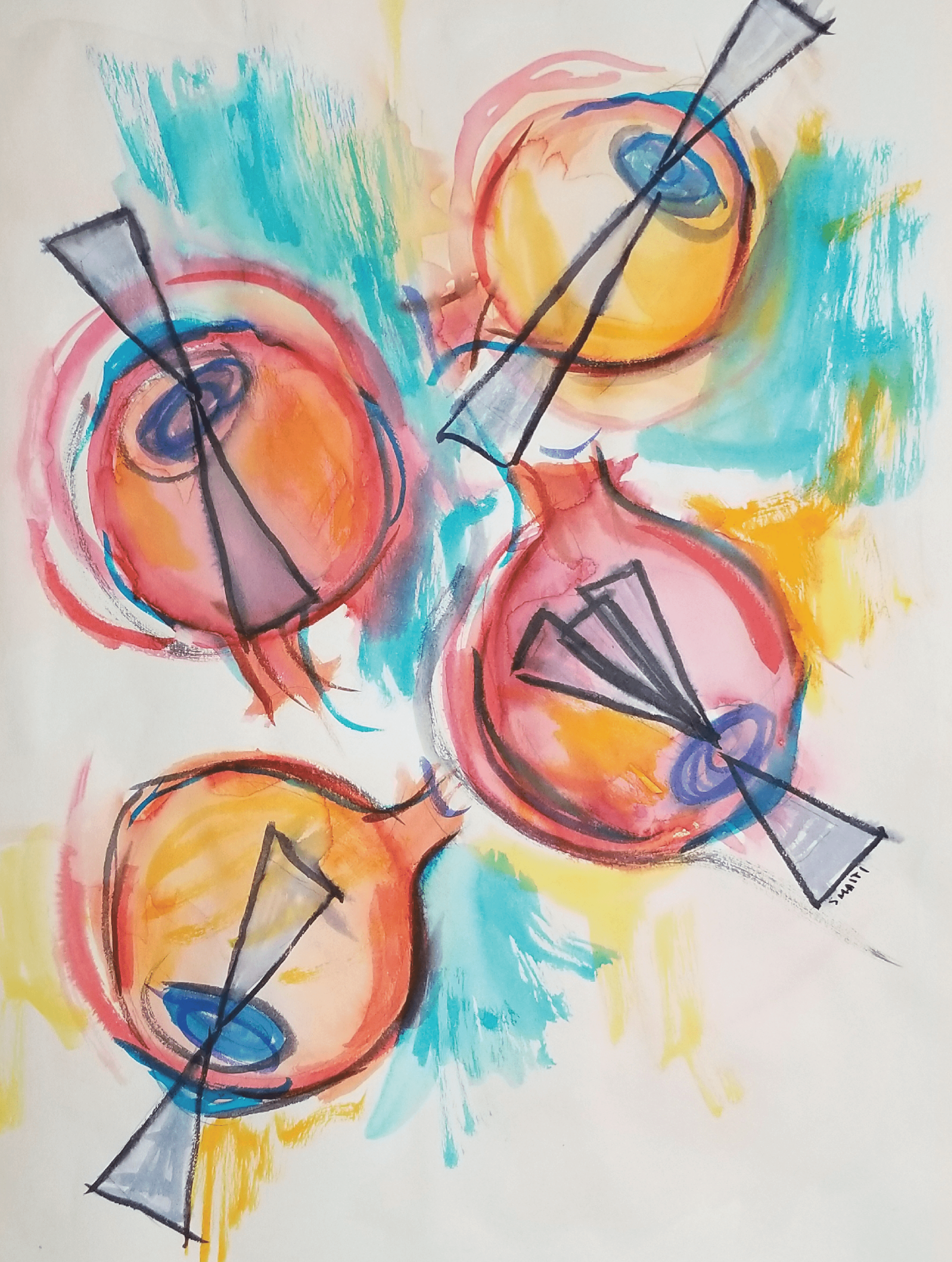
Sathi Maiti is an ocular surface disease clinical research fellow at the Periman Eye Institute and primary care optometrist in Seattle, Washington, USA. She completed her undergraduate degree in molecular, cellular, and developmental biology at the University of Washington in 2009 and her Doctorate of Optometry at UC Berkeley in 2014. She has done lymphatic and genetics research, taught cell biology and human anatomy, and has always been inspired by the forms found in microscopy, biology and the natural world. While she never formally studied fine art after high school, she has been making art as a hobby for her entire life.
She says: “I enjoy experimenting with different mediums from traditional drawing and painting to collage, embroidery and other fiber art. I gain inspiration from the world around me, and as an eye doctor that has come to include a lot of eyeballs! We are very lucky as eye care practitioners to work with a structure as complex, physiologically fascinating, and aesthetically interesting as the eye.”
Maiti shares her art and other eye-related content on her instagram account @dr.maitiseyeballsandstuff
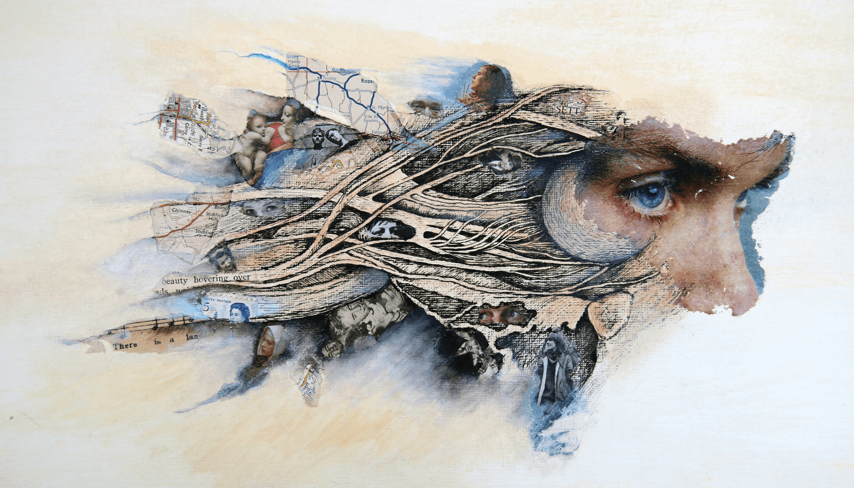
Jennie Jewitt-Harris, featured prominently in last year’s Art of Eyes, is a CEO of the Transplant Links charity, artist and physician. She has a PhD in Fine Art for her research into the reasons why many people refuse to donate their corneas after death. Her exhibition, “Anything But the Eyes,” will be presented at South Hill Park Mirror Gallery in Bracknell, UK, from July 8 to October 2, 2021.
This year, she has shared her new work with The Ophthalmologist. Can you tell whose eyes are featured in Anatomy vs Psychology?
Denice Barsness is Technical Director at the Ophthalmic Diagnostic Center at CPMC Department of Ophthalmology in San Francisco, California, USA. She is very experienced in all aspects of ophthalmic assisting and technology, as well as imaging. She has over 25 years of experience as faculty staff member of ophthalmic imaging education institutions and is Past President of Ophthalmic Photographers’ Society.
A. David Flug is a board-certified ophthalmologist who has practiced ophthalmology and eye surgery in Forest Hills, New York, USA, since 1978. He is a graduate of the Hahnemann Medical College and did an ophthalmology residency at the Long Island Jewish Medical Center. He is currently on the teaching staff of the North Shore University, Long Island Jewish Medical Center and on staff at Flushing Hospital, and the renowned Mackool Eye Institute.
Christina Appleman is a Certified Ophthalmic Medical Technologist and an Ophthalmic Research Technician in Rockville, Maryland, USA.
Of her art, she says: “My grandfather used to make wine as a hobby so there were always wine corks laying around that were begging to be utilized. I enjoy coming up with creative and unique ways to upcycle the corks.”
Appleman’s work can be found on Instagram @thewineingtwins.