Lowering intraocular pressure (IOP) remains the cornerstone of glaucoma treatment, yet retinal ganglion cell (RGC) loss can continue even when pressure is adequately controlled. New preclinical research points to impaired cellular waste disposal – and, specifically, the failure to clear damaged mitochondria – as a potential contributor to this continuing neurodegeneration.
In a study published in Molecular Neurodegeneration, Prabhavathi Maddineni and colleagues investigated whether defective autophagy and mitophagy contribute to mitochondrial accumulation, oxidative stress, and RGC degeneration in glaucoma. The team used glucocorticoid-induced and myocilin-associated mouse models of glaucoma, alongside mitophagy reporter mice, RGC-specific genetic manipulation, and ex vivo human retinal tissue.
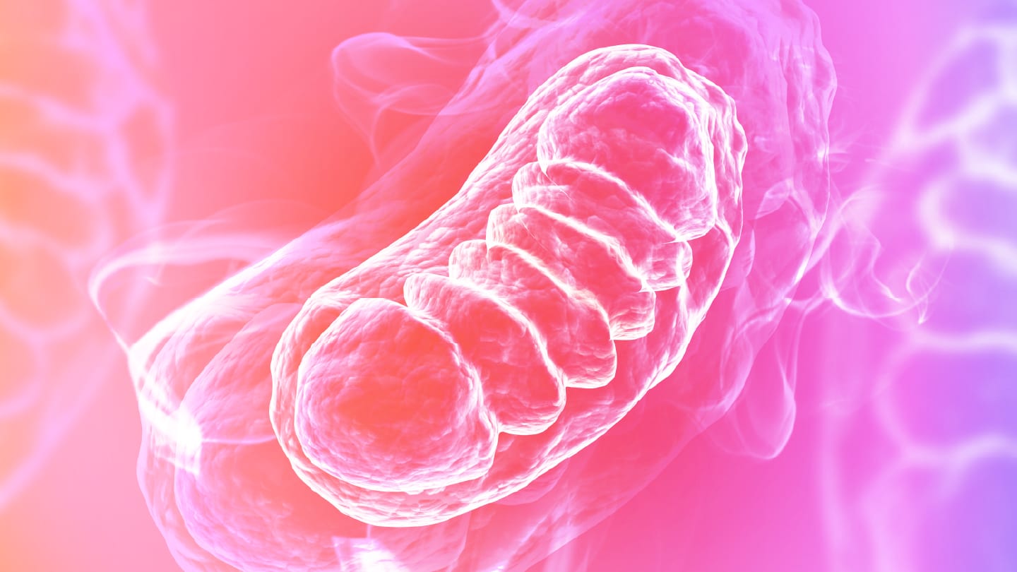
The researchers – based at the University of Missouri, the University of California, and the University of North Texas Health Science Center – first found that sustained IOP elevation was associated with an accumulation of structurally abnormal mitochondria in RGCs and optic nerve axons. Transmission electron microscopy revealed swollen mitochondria with disorganized or fragmented cristae, while retinal tissue showed increased markers of mitochondrial accumulation and oxidative DNA damage.
These mitochondrial changes did not appear to be simply a consequence of advanced degeneration. Using Mt-Keima reporter mice, which allow mitophagy flux to be tracked, the researchers detected significantly reduced mitochondrial clearance after five weeks of ocular hypertension – before detectable RGC loss and axonal degeneration. Impaired mitophagy persisted once neurodegeneration was established.
To test whether defective autophagy might itself drive neurodegeneration, the researchers selectively disrupted the autophagy-related gene Atg5 in RGCs. Six weeks later, the treated eyes showed abnormal mitochondrial accumulation, oxidative DNA damage, impaired RGC function, approximately 39% RGC loss, and around 59% loss of healthy optic nerve axons.
The team then asked whether restoring autophagy might be protective. Intravitreal treatment with Torin 2 – an mTOR inhibitor that activates autophagy – increased mitophagy and preserved RGC function in the glucocorticoid-induced glaucoma model without altering IOP. Mean pattern electroretinography amplitudes were 31.82 µV in Torin 2-treated hypertensive eyes, compared with 17.36 µV in control-treated hypertensive eyes. The treatment was also associated with a 37% increase in RGC number and a 51% increase in morphologically intact axons.
These findings were extended beyond mouse tissue. In an ex vivo human retinal explant model cultured under neurotrophic factor-deprived conditions, Torin 2 was found to increase RGC survival by approximately 49.7%.
The work remains preclinical, and in the myocilin model some neuroprotection may have resulted from an accompanying reduction in IOP. Torin 2 itself is also unlikely to provide the final therapeutic answer. Its broad inhibition of mTOR may complicate translation, with the authors noting that their “future studies will focus on more selective autophagy/mitophagy modulators or pathway-specific interventions to validate and refine mitophagy-targeted therapies for glaucoma.”
Even so, the study places defective mitochondrial clearance upstream of RGC degeneration and raises the possibility of targeting autophagy as an IOP-independent neuroprotective strategy – potentially addressing one of glaucoma care’s most persistent unmet needs.