
Since its introduction, Heidelberg Engineering’s SPECTRALIS platform has been a benchmark in multimodal retinal imaging – trusted in clinics, hospitals, and research centers around the world. With the upcoming new release on SPECTRALIS, the company takes the next step in imaging evolution: enhancing workflow efficiency, imaging speed, and diagnostic confidence. This next expansion of the SPECTRALIS multimodal functionality adds a suite of new tools – from Green Autofluorescence to faster OCT Angiography Module and flexible reporting options – that build on the platform’s well-established strengths in enhancing diagnostic confidence and continuously improving patient care – exclusively compatible with HEYEX 2.
Faster and personalized imaging
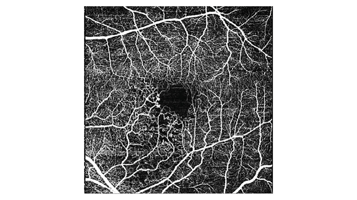
The new SPECTRALIS software gives clinicians more control to tailor imaging protocols through the addition of the SHIFT 250 kHz OCTA scan speed. Joining the existing 20 kHz, 85 kHz, and 125 kHz speeds, it allows users to prioritize either faster acquisition or maximum detail – depending on the clinical setting and patient needs. It also introduces DART (Dense Automatic Real Time) OCTA imaging, which applies ART averaging of up to 100 B-scans to deliver high-detail, noise-reduced images of retinal vasculature. Noise reduction allows for improved image quality, ensuring small details aren’t missed during examination. These developments are supported by enhanced TruTrack Active Eye Tracking, which now offers faster and more responsive motion compensation. By stabilizing images during both extended and high-speed scans, the system maintains alignment and consistency without the need for hardware adjustments – helping ensure reliable image quality throughout the exam.
Confident diagnostic decisions
The upcoming release also broadens the diagnostic scope of SPECTRALIS through a range of new imaging capabilities designed to provide deeper insights into retinal and optic nerve health.
Green Autofluorescence Module
Using 518 nm excitation, the Green Autofluorescence Module improves visualization of foveal sparing in macular atrophy and supports the assessment of lipofuscin distribution and retinal pigment epithelium loss – valuable in managing conditions such as geographic atrophy (GA) and Stargardt’s disease. In combination with BluePeak blue laser autofluorescence, it strengthens the multimodal imaging portfolio for assessing macular atrophy.

Low Luminance Autofluorescence
For light-sensitive patients or those with advanced retinal degeneration, the new low-luminance mode allows clinicians to select blue laser power for reflectance and autofluorescence imaging between 25% and 100%. This minimizes light exposure and can help improve patient comfort throughout the examination.
Change Analysis on Glaucoma Module Premium Edition (GMPE)
Glaucoma assessment is strengthened through the new Posterior Pole Change Analysis, which offers the flexibility to combine or segment layers in the Ganglion Cell Complex (GCC) and enables visualization of patterns of structural change in the macular over time, in one view, on one screen. Users can also freely select their desired posterior pole scan as baseline to track changes or response to treatment intervention.
Faster scans, improved comfort
Higher scan speed of 250 kHz together with enhanced active eye tracking for OCTA shortens acquisition time and thus may support efficient clinical workflows and improve patient comfort. Shorter examination times benefit patients who struggle to maintain fixation – particularly elderly, pediatric, or low-vision individuals.
Low Luminance Autofluorescence ensures that advanced diagnostics are accessible even for those who are light-sensitive or require less than standard laser power based on their retinal condition.
Flexible and personalized workflows
Beyond imaging performance, the new software focuses on practical workflow improvements that help clinicians work more efficiently.
Custom scan presets
Users can create additional custom scan presets directly in the acquisition module. Each preset stores defined parameters, allowing teams to standardize imaging protocols across clinics, research sites, or patient groups.
Multiselect report printing
The multiselect print function enables multiple reports to be generated or merged into a single PDF in one step. Saved as templates, these configurations simplify documentation, reduce administrative time, and support consistent reporting practices.
Heidelberg Engineering continues to focus on ensuring that technology adapts to clinical workflow.
Confident diagnostics and personalized patient care
This next release on SPECTRALIS reflects an ongoing evolution in ophthalmic imaging – ensuring fast, reliable, and patient-centered diagnostics. With enhanced speed, advanced clinical modules, and streamlined workflow tools, it brings high-performance imaging into everyday practice.
Heidelberg Engineering pioneers imaging and data technologies to optimize ophthalmic solutions and support learning that advances research and empowers eyecare professionals who want to improve the holistic health of patients.
By supporting both clinical and research environments, the upcoming software continues to strengthen the SPECTRALIS platform as a trusted foundation for outcome-based, multimodal eye care. It represents a steady progression toward imaging that is faster, smarter, and more adaptable – designed to meet the needs of today’s ophthalmology and tomorrow’s innovations.
The software is currently not available for clinical use. Please note that not all product features and modalities described will be available for sale in all countries.
