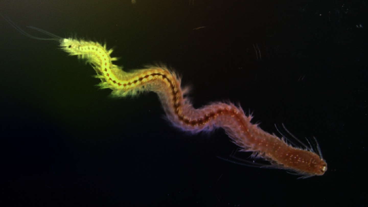
When ophthalmologists look for inspiration on retinal neurogenesis, Platynereis dumerilii — a small marine bristleworm — is not necessarily the first organism that springs to mind. But a new study in Nature Communications positions this unassuming annelid as an unexpectedly powerful model for understanding adult retinal plasticity, stem cell dynamics, and even the evolutionary logic of eyes.
Using single-cell RNA sequencing, a research group based at the Department of Neurosciences and Developmental Biology, University of Vienna, Austria, dissected the cellular organization of the adult Platynereis head — focusing on its sophisticated camera-type eyes, which strikingly echo the architecture of vertebrate and cephalopod eyes. Their analyses uncovered a previously unrecognized stem-cell niche at the rim of the cup-shaped retina, functionally reminiscent of the vertebrate ciliary marginal zone (CMZ). This zone continuously seeds new photoreceptor and support cells — fueling lifelong ocular growth.
High-resolution EdU pulse-chase experiments showed that proliferating cells localize to a distinct ring abutting the glass-body “lens” — precisely where radial arrays of new photoreceptors appear to emerge. In short, this simple worm builds and maintains its retina using a blueprint astonishingly parallel to vertebrate systems.
Wild-type worms raised under light–dark cycles showed robust EdU incorporation in the eye; those kept in darkness did not. C-opsin1 mutants lost this light-dependent proliferative response, and their retinas lacked a subset of mature photoreceptors — suggesting impaired differentiation.
For ophthalmologists, the parallel is tantalizing: light-responsive regulation of retinal stem cells — long documented in fish — emerges here in an evolutionarily distant invertebrate.
The study also identified a striking, whole-brain transition during reproductive maturation: a global shutdown of proliferation accompanied by a molecular shift towards adult neural stem-cell quiescence.
This coexistence of neurogenic and quiescent signatures — validated in vivo by HCR imaging — echoes mammalian aNSC biology, but in a system that naturally undergoes a terminal, semelparous reproductive transition.
While Platynereis is far from the clinic, its retina may help illuminate fundamental rules governing stem cell behavior — rules that continue to shape how ophthalmology approaches regeneration, repair, and retinal engineering.
