During early medical training, it’s vital for all doctors – especially ophthalmologists – to gain a basic grounding in human head, neck and orbital anatomy. But by the time those same doctors earn their specialist credentials in clinical ophthalmology, their years of examining cadaveric remains are often far behind them. Unfortunately for advanced trainees who want to refresh their knowledge, access to specimens is often restricted or otherwise difficult – most facilities are centralized in larger cities, while others may have limited open hours or be available only to medical students at the host institution. In more remote locations, the task becomes even more challenging; many locations may not have access to cadaveric specimens at all due to health and safety regulations, cultural or ethical considerations, lack of necessary storage requirements, or the prohibitive cost of cadaver bequest programs. As a result, many trainee ophthalmologists are limited to photographs and diagrams when updating and increasing their knowledge of orbit, head and neck anatomy – a distinct disadvantage in the study of a region with such complex 3D anatomy.
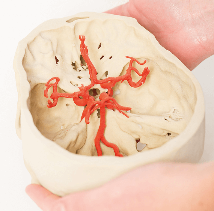
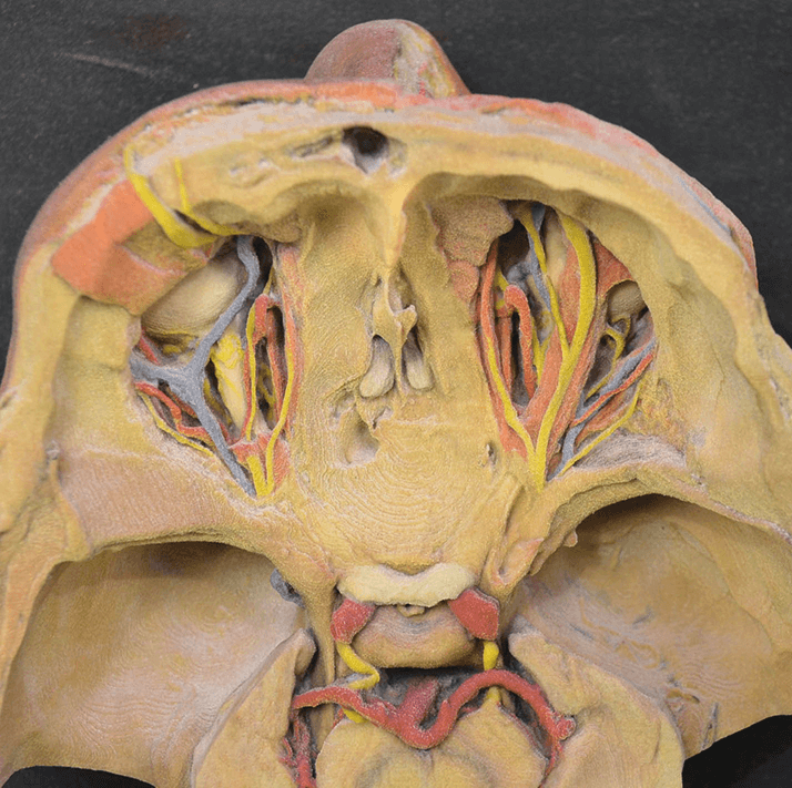
How can these difficulties be addressed? A group from Monash University (Clayton, Victoria, Australia) is exploring 3D printing as a new and inexpensive way to tackle them (1). Historically, single-color 3D printouts have been of limited value in soft tissue anatomy, because color provides important information. But recent developments in full-color and multiproperty 3D printing are beginning to overcome this obstacle, making it possible to create low-cost, high-resolution reproductions of prosected human cadaver orbits. The group began by preparing teaching prosections of the orbit from a superior, a lateral and a medial perspective, optimizing the number of features displayed in each. Once the prosections were complete, researchers created surface meshes of the specimens using a high-resolution 3D scanner that captured both shape and color details. For added value, they also enhanced the colors of important features using digital painting, to ensure that vessels and nerves that look similar in wet prosections were easier to see in the 3D reproductions. The resulting prints were found to be highly realistic and clearly represented the most delicate structures of the orbit. Not only that, but they offer a number of advantages over either cadaver or plastinated specimens – accuracy, detail, rapid reproduction, easy storage, portability, and the avoidance of cultural, ethical and safety concerns associated with human cadaveric material. There’s promise for future developments, too; though the output is only as good as the input (in this case, the prosections), the investigators have also managed to create 3D printouts from MRI and CT datasets. Because of their many advantages, the printouts offer an excellent substitute where cadaveric material isn’t available – and can serve as a useful adjunct even where it is.
References
- JW Adams et al., “3D printed reproductions of orbital dissections: a novel mode of visualising anatomy for trainees in ophthalmology or optometry”, Br J Ophthalmol, [Epub ahead of print] (2015). PMID: 25689987.
