Richard C. Allen
Patients who have an anophthalmic socket can have a variety of issues to contend with. It is important to remember that they have lost one of their vital organs, leaving them with monocular vision and depth perception difficulties. More importantly, they no longer have a “spare tire” that can be utilized should their remaining eye also fail them, and so it’s crucial to ensure this remaining eye stays healthy. A secondary concern for an ophthalmologist, but often the primary concern of the patient, is the cosmetic outcome after enucleation or evisceration.
In the care of a patient with an anophthalmic socket, attention is directed to three main points: the surface area of the conjunctiva, the volume in the socket, and the motility of the ocular prosthesis. All of these factors can affect the external appearance of the patient. Volume is affected by the size of the ocular implant that has been placed – the larger the implant, the thinner the prosthesis, which in turn results in a better fitting, better moving prosthesis, and a more symmetrical external appearance. Motility is affected by many factors, including the fit of the prosthesis, the type of implant placed, and the surface area of the conjunctiva. The most complex problem that can occur during the procedure is generally a loss of surface area of the conjunctiva (contraction), which can result in an inability to retain the prosthesis, apparent lid malposition (lagophthalmos, retraction), loss of the fornices, and loss of motility of the prosthesis.
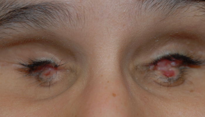
When a patient presents with contraction, it should first be determined if the contraction is segmental or global. Segmental contraction includes scar bands and loss of the upper or lower fornix, while global contraction refers to diffuse shortening. The etiology of the contraction needs also to be ascertained. This can be present at the primary surgery in which the conjunctiva was already compromised due to autoimmune disease or multiple previous surgeries. There may be a predisposition to contracture due to previous radiation to the orbit or chemotherapy (See Figure 1).
There are also a number of acquired processes which can cause socket contraction, including chronic infection or inflammation due to conjunctivitis or nasolacrimal duct obstruction, an underlying inflammatory condition, previous repairs of an exposed orbital implant, or conjunctival malignancy.
If the contraction of the conjunctiva cannot be explained, a biopsy of the conjunctiva should be performed. The specimen should be sent for anatomic pathology – as well as direct immunofluorescence – to look for processes such as mucous membrane pemphigoid and lichen planus.
Malignancy can also cause contracture of the conjunctiva, and this should always be suspected and ruled out with biopsy if the contraction cannot be explained. It is postulated that chronic inflammation of the anophthalmic socket predisposes the patient to conjunctival malignancy (1). In addition, if previous radiation has been applied to the area, this can increase the risk of a malignancy. Conjunctival squamous cell carcinoma is the most common malignancy reported in anophthalmic sockets, but there have also been reports of sebaceous carcinoma and melanoma for such patients.
Treatment of the contracted socket should depend on the etiology and the extent of the contraction. If there is mild contraction thought to be due to socket inflammation, the prosthesis should be refitted, and basic hygiene maintenance procedures of the anophthalmic socket explained to the patient.
Among anophthalmic patients, there is a common misconception that their prosthesis needs to be taken out frequently and cleaned; however, the less the patient manipulates the prosthesis, the better they usually do. While the prosthesis should not be taken out frequently, it should be polished every six-twelve months by the ocularist. Preservative-free lubricants should be used as needed. In patients with mild contraction, the conjunctiva should be examined closely to check for a progressive cicatricial process. The contralateral side should also be examined. Lastly, the inflammation can be treated with topical or local (injection) agents. Topical agents usually consist of a short course (one month) of steroids. Topical 5-fluorouracil (5-FU) and mitomycin have also been described, and, more recently, injections of 5-FU under the conjunctiva have been investigated, with successful outcomes demonstrated (2).

Figure 1: 10-year-old patient with significant bilateral anophthalmic socket contraction. The patient has a history of bilateral retinoblastoma, s/p bilateral enucleation followed by radiation to the orbits.
An early sign of socket contracture is marginal entropion and lash ptosis. This can be treated with a tarsal fracture, but only if there is no evidence of a progressive process. Another treatment – pressure conformers – have also been promoted by some specialists, with the idea that pressure on the surface of the conjunctival acts as a tissue expander.
If there is focal or segmental contraction, this can be treated with adhesiolysis alone, or else with placement of an amniotic membrane graft or a mucous membrane graft. Generally, split or full-thickness skin grafts should be avoided in anophthalmic sockets – the production of keratin from the skin results in a foul discharge.
The lower fornix is a commonly affected area of shortening that results in difficulty retaining the prosthesis. The anatomical change that anophthalmic sockets go through with time may also affect the loss of the lower fornix. Many treatments have been documented in previous literature, including placement of fornix sutures and reconstruction of the fornix with multiple different grafts and flaps (3).
In my experience, the most difficult socket contraction to treat is severe global or recurrent contraction. This is common in a socket that has a history of multiple surgeries or previous radiation. It is usually a progressive process and may have a component of poor vasculature that is impeding the survival of free grafts. In patients who have relatively healthy conjunctiva but contraction due to previous conjunctival surgeries usually related to implant exposure, removal of the implant and replacement with a dermis fat graft is an effective procedure. The dermis fat graft serves two purposes: to provide volume to the socket and to expand the conjunctival surface (4).
For patients who have a global conjunctival shortage, a mucous membrane graft needs to be performed. With significant contraction, a conformer wrapped with mucous membrane works well for the graft. However, if the socket is thought to be relatively avascular due to multiple previous surgeries or radiation, vascularized tissue needs to be moved into the socket. This can be accomplished with a free flap (performed in conjunction with a microvascular surgeon) or a local flap. Common local flaps to use around the orbit include temporalis muscle, a temporoparietal flap, and a buried, de-epithelialized mid-forehead flap. In patients with previous failures that are undergoing extensive reconstructions, it is often advantageous to place a tarsorrhaphy for three-four months with a conformer.
In summary, while patients with socket contraction can often be very difficult to treat, having a systematic approach in place and accurately determining the etiology of the contraction can often produce satisfactory results for the patient.
References
- P Bonavolontà et al., “Conjunctival Carcinomas Arising in the Anophthalmic Socket,” J Craniofac Surg, 32, 2 (2021). PMID: 33705043.
- S Kamal et al., “Serial sub-conjunctival 5-Fluorouracil for early recurrent anophthalmic contracted socket,” Graefes Arch Clin Exp Ophthalmol, 12, 2797 (2013). PMID: 24132696.
- K Vahdani, GE Rose, “Long-term Outcome of Conjunctival Transposition Flaps for Contracted Sockets,” Ophthalmic Plast Reconstr Surg, [Online ahead of print] (2024). PMID: 39158478.
- N Jovanovic et al., “Reconstruction of the Orbit and Anophthalmic Socket Using the Dermis Fat Graft: A Major Review,” Ophthalmic Plast Reconstr Surg, 6, 529 (2020). PMID: 32134765.
