Managing patients with retinal disease can be challenging – they present to practices with a greater diversity of age and disease states (and in far greater numbers) than ever before. As always, effective initial disease detection and progression management – with the right treatment option at the right time – remains crucial for truly effective long-term vision preservation. And this is where widefield imaging comes into play. Widefield imaging has shown us the possibility of uncovering more pathology by imaging a larger area of the retina than standard fields. Widefield imaging allows for more thorough documentation and detection of peripheral retinal pathology, leading to earlier diagnosis of diseases and better management of patients. However, traditional fundus imaging systems remain the gold standard for macular disease diagnosis and optic nerve evaluation. The launch of ZEISS HD ultra-widefield system represents the next generation of widefield fundus imaging. Leveraging ZEISS optics, the technology promises to deliver exceptional clarity from the macula to the far periphery in a true color image. True color images are generated through sequential illumination by a full spectrum red, green and blue LEDs. Unlike other scanning laser technologies, ZEISS HD-UWF images closely resemble the coloration of the fundus as seen through direct observation. In addition to imaging the peripheral retina in high resolution, the ZEISS HD UWF system captures high quality images of the optic nerve head and the macula. This eliminates the need for clinicians to maintain more than one fundus imaging system in their practice and helps them manage a broader range of patients.
Diabetic retinopathy
Ultra-widefield (UWF) imaging is the ideal tool to detect diabetic retinopathy. Recent studies have demonstrated the importance of peripheral retinopathy, and its role in predicting disease progression. UWF imaging provides a fast, reliable method for imaging the peripheral retina. Green channel separation images further highlight hemorrhages and microaneurysms, making it easier to visualize subtle retinopathy. To achieve an ultra-wide field of view, many systems compromise on image resolution. The ZEISS system maintains excellent clarity and color, giving the ability to zoom into any part of the image and view the retina in great detail. In this case study, the patient has neovascularization nasal to the optic disc. The fine neovascular frond can be seen clearly on the image. Having both field of view and good resolution gives the clinician confidence when managing their diabetic patients.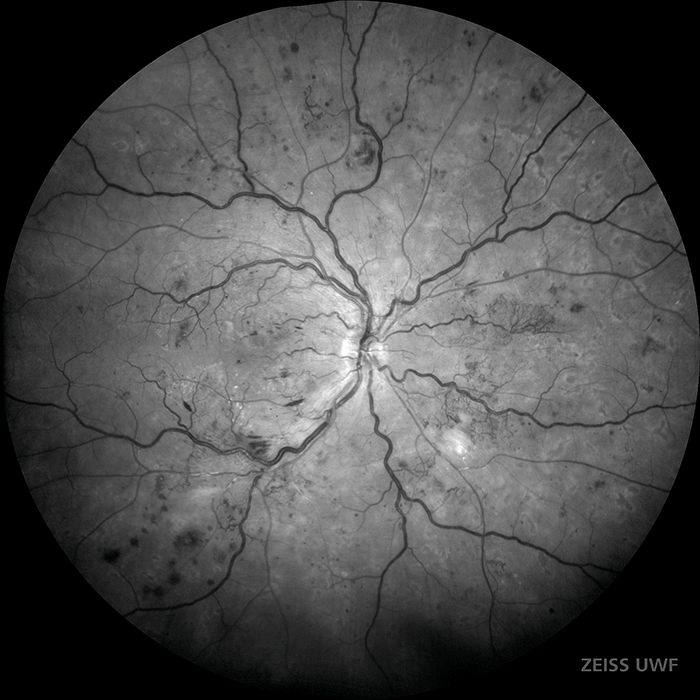
AMD
For dry age-related macular degeneration, accurate coloration is important for documenting RPE pigment changes and drusen. This patient was imaged on the ZEISS UWF system as part of his exam. Scattered drusen can be seen throughout the macular region. Reticular degeneration, which is a risk factor for AMD progression, can also be seen in the peripheral retina. Even though AMD is primarily a macular disease, UWF imaging still plays a role in documenting peripheral risk factors. The ZEISS system has the advantage of peripheral imaging, while still maintaining the image quality of a traditional fundus camera.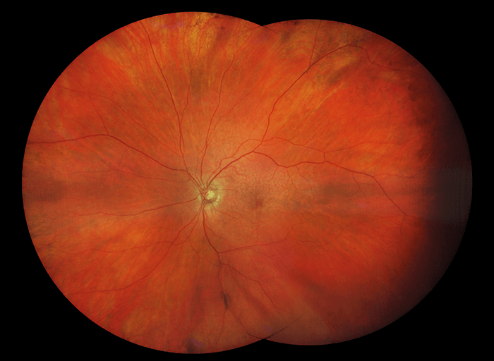
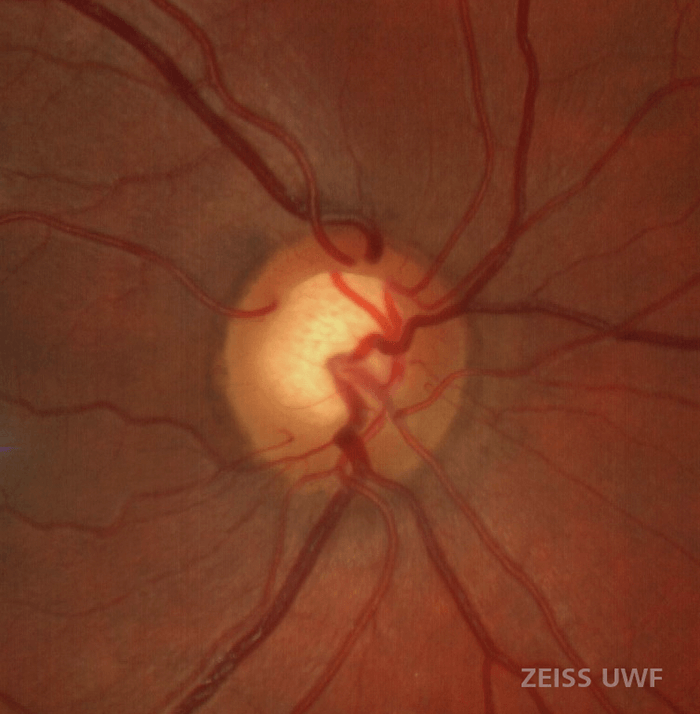
Glaucoma
Because of the subtle and slow-changing nature of the disease, glaucoma necessitates precise, high resolution, repeatable documentation of the optic disc, whether it be with OCT, fundus photos, or visual fields. In fundus imaging, having a high quality, true color image that allows for quantitative measurements is critical. ZEISS color and resolution enables evaluating focal changes in rim tissue or nerve pallor with confidence. The review software makes it easy to compare images over time, aiding in the detection of early glaucomatous changes.