Conexiant
Login
  • Corneal Physician
  • Glaucoma Physician
  • New Retinal Physician
  • Ophthalmology Management
  • Ophthalmic Professional
  • Presbyopia Physician
  • Retinal Physician
The Ophthalmologist
  • Explore

    Explore

    • Latest
    • Insights
    • Case Studies
    • Opinion & Personal Narratives
    • Research & Innovations
    • Product Profiles

    Featured Topics

    • Anterior Segment
    • Glaucoma
    • Retina

    Issues

    • Latest Issue
    • Archive
  • Subspecialties
    • Cataract
    • Cornea
    • Glaucoma
    • Neuro-ophthalmology
    • Oculoplastics
    • Pediatric
    • Retina
  • Business

    Business & Profession

    • Professional Development
    • Business and Entrepreneurship
    • Practice Management
    • Health Economics & Policy
  • Training & Education

    Career Development

    • Professional Development
    • Career Pathways

    Events

    • Webinars
    • Live Events
  • Events
    • Live Events
    • Webinars
  • Community

    People & Profiles

    • Power List
    • Voices in the Community
    • Authors & Contributors
  • Multimedia
    • Video
    • Podcasts
Subscribe
Subscribe

False

Advertisement
The Ophthalmologist / Issues / 2018 / Jun / Ultra-High Fidelity Fundus
Retina Sponsored

Ultra-High Fidelity Fundus

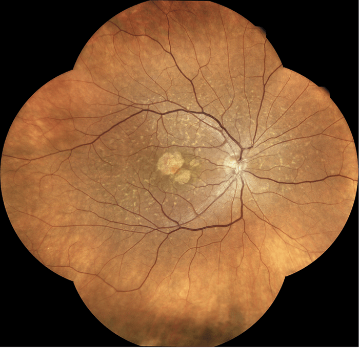
Imagine combining the best features of scanning laser ophthalmoscopy and standard fundus photography to give retinal images that are both high-resolution and wide field. Dr. Valentina Sarao of the University of Udine, Italy, shares her experience of the EIDON TrueColor Confocal Scanner.

Sponsored By CENTERVUE 6/27/2018 1 min read

Share

Figure 1. 110° Mosaic of retina in a patient with Stargardt disease, as imaged by the EIDON TrueColor Confocal Scanner.

What are the main features of EIDON?
Uniquely, EIDON offers both confocal technology and white light illumination in a single system. The resulting high resolution fundus images – displaying actual retinal colors – greatly improve diagnosis and monitoring of retinal disease. Also, the confocal capability permits high-resolution, high-contrast images even in cataractous eyes – where conventional flash fundus cameras are of limited use. In addition, patients often dislike the flash, whereas EIDON’s soft light-source is more comfortable and requires no pupil dilatation. Notably, EIDON permits a remarkably wide field of view (Figure 1). A single exposure captures 60° with a viewing angle of up to 110° in automatic-mosaic mode; manual operation can generate even wider montages without losing resolution. EIDON’s operation is highly intuitive, making for a brief learning curve; automatic alignment of pupil and retina means that minimal operator involvement is required. Finally, the system is very versatile; users can switch between automatic alignment and manual joystick mode as required. Thus, physicians can customize its operation to capture specific pathologies.

How does EIDON benefit your clinic?
In my clinic, EIDON is a standard – and essential – diagnostic technique for posterior segment disease; it simplifies capturing the comprehensive retinal documentation required for all patients. It discriminates between retinal hemorrhages and microaneurysms in eyes affected by diabetic retinopathy; it reveals epiretinal membranes that traditional fundus cameras miss; and its fundus autofluorescence capability is key for evaluating retinal dystrophies, RPE abnormalities and geographic atrophy. Furthermore, EIDON’s flexible operation supports multifield acquisitions (non-standard fields may be selected by displacing the internal fixation target), and it is very fast.  EIDON’s high level of automation makes it accessible to a broad range of personnel, including optometrists and technicians; it is therefore easy to introduce into daily practice. Importantly, EIDON provides high resolution images of both macula and retinal periphery – essential for diagnosing severe retinal conditions. I find the high-resolution display tablet invaluable for sharing information with patients or colleagues, and for training residents.
How will EIDON be used in the future?
The unique combination of confocal technology and white light illumination, providing high-resolution, TrueColor retinal images, should make EIDON essential in today’s ophthalmology practice. I see it becoming a standard primary care tool in screening for diabetic retinopathy, glaucoma and AMD.

Related Content

Newsletters

Receive the latest Ophthalmology news, personalities, education, and career development – weekly to your inbox.

Newsletter Signup Image

False

Advertisement

False

Advertisement

Explore More in Ophthalmology

Dive deeper into the world of Ophthalmology. Explore the latest articles, case studies, expert insights, and groundbreaking research.

False

Advertisement
The Ophthalmologist
Subscribe

About

  • About Us
  • Work at Conexiant Europe
  • Terms and Conditions
  • Privacy Policy
  • Advertise With Us
  • Contact Us

Copyright © 2025 Texere Publishing Limited (trading as Conexiant), with registered number 08113419 whose registered office is at Booths No. 1, Booths Park, Chelford Road, Knutsford, England, WA16 8GS.

Disclaimer

The Ophthalmologist website is intended solely for the eyes of healthcare professionals. Please confirm below: