Subscribe to Newsletter
Subspecialties Cataract, Refractive

Two IN One

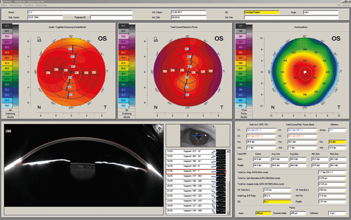
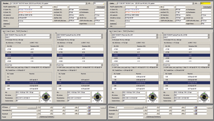
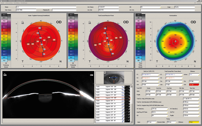
All surgeons want the best for their patients, and when it comes to refractive outcomes, anterior segment analysis plays a key role. The Pentacam® and Pentacam® HR provide a gold-standard in anterior segment tomography. Now meet the newest member of the family – the Pentacam® AXL.

Based on the trusted Pentacam® HR system, which offers high-resolution Scheimpflug imaging, the Pentacam® AXL offers the additional feature of axial length measurement – a crucial part of accurate IOL calculation and customized IOL selection. Through partial coherence interferometry biometry, the Pentacam® AXL collects several successive axial length measurements; patient eye movement is detected by the pupil camera and corrected for, and a 3D model of the anterior segment based on ray tracing allows for the correction of individual optical distortions. Using fourth generation IOL formulas, the system can automatically calculate IOL power. Because the total corneal refractive power (TCRP) map shows the influence of the posterior corneal surface with regard to total corneal astigmatism axis, magnitude and regularity, the IOL calculation software can account for posterior corneal astigmatism. The software also accounts for prior refractive surgery and pre-existing conditions, ensuring a reliable IOL power calculation for any IOL type in both virgin and post-refractive eyes.

The upshot? The Pentacam® AXL enables full screening before corneal refractive and cataract procedures. Laser vision correction procedures can be planned effectively; patients can be screened for existing diseases such as Fuchs endothelial dystrophy or angle-closure glaucoma; prior refractive surgeries can be detected; and crystal lens densitometry can be used to plan femtosecond laser-assisted cataract surgery (FLACS). Now, accurate IOL calculation can also be achieved – as well as customized IOL selection regarding multifocal, toric and aspheric IOLs, all from one device.

Figure 1. TCRP map for the right eye.

Figure 2. TCRP map for the left eye.

Figure 3. IOL calculations.

Pentacam® AXL in Action

A case study by Ina Conrad-Hengerer, MD, University of Heidelberg, Germany.

A 63-year-old woman presented with subjective loss of visual acuity and increased glare. Corrected distance visual acuity (CDVA) was reduced in both eyes; 20/63 (-0.5 D -1.0 D × 98°) in the right eye and 20/40 (-1.0 D -0.75 D × 75°) in the left eye. Examination of the anterior segments revealed senile cataract without corneal pathology. Before medical mydriasis or further examinations, Scheimpflug imaging and anterior segment analysis by Pentacam® AXL was performed. The postoperative target refraction was emmetropia for distance, and fundoscopy showed no macular changes.

The TCRP map showed increased corneal astigmatism of the axial topography from 0.9–1.6 D (right eye) and from 1.4–1.8 D for the left eye, respectively (Figures 1 and 2). Usually, toric IOL implantations are considered based on spectacle correction and anterior corneal keratometry values – but that approach would not have identified this result. Total spherical corneal aberrations (6 mm zone) were 0.353 µm (right eye) and 0.305 µm (left eye), revealing that an asphericity-correcting IOL could be a good option. Total corneal irregular astigmatism (4 mm zone) was 0.154 µm (right eye) and 0.145 µm (left eye); as 0.3 µm is the recommended limit to avoid photic phenomena, multifocal IOLs could be offered to this patient.

The IOL calculations are presented in Figure 3. Femtosecond laser-assisted cataract surgery followed by implantation of monofocal aspheric toric IOLs was performed (right eye, +18.0 D 1.5 D/6°; left eye, +18.0 D 2.25 D/6°). One day after surgery, UDVA was 20/20 in both eyes, increasing to a UCVA of 20/16 one month after surgery. Objective refraction measured by Nidek AR310A was +0.25 D  0.25 D/110° (right eye) and plano -0.25/60° (left eye).

www.pentacam.com

Receive content, products, events as well as relevant industry updates from The Ophthalmologist and its sponsors.

When you click “Subscribe” we will email you a link, which you must click to verify the email address above and activate your subscription. If you do not receive this email, please contact us at [email protected].
If you wish to unsubscribe, you can update your preferences at any point.

Related Case Study
Uncovering Ocular Comorbidity

| Contributed by Quidel

Related Product Profiles
Sulcus-based enhancement of visual quality

| Contributed by Medicontur Medical Engineering Ltd

Less Steps, More Vision

| Contributed by Medicontur Medical Engineering Ltd

Product Profiles

Access our product directory to see the latest products and services from our industry partners

Here
Most Popular
Register to The Ophthalmologist

Register to access our FREE online portfolio, request the magazine in print and manage your preferences.

You will benefit from:
  • Unlimited access to ALL articles
  • News, interviews & opinions from leading industry experts
  • Receive print (and PDF) copies of The Ophthalmologist magazine

Register

Disclaimer

The Ophthalmologist website is intended solely for the eyes of healthcare professionals. Please confirm below: