It is hard to underestimate the contribution OCT has made to eyecare. It has transformed our understanding of the pathology and pathophysiology of the eye – particularly, the retina, but also the cornea. OCT has made giant strides over the years – the main example being the change from time-domain to spectral-domain OCT, which heralded a huge increase in image quality, boosting utility of the technology. Another big stride forward appears to be on the horizon – and it all boils down to better speckle removal. OCT relies on the coherent detection of backscattered light to image tissue morphology, which comes with an intrinsic drawback: speckle noise, arising from the interference of light scattered from multiple points within the tissue being examined. These scattered photons can cancel each other out, giving a false-black speckle on the image; or they can reinforce each other, generating a false-white speckle. In either scenario, these are artifacts: unhelpful and unwanted.
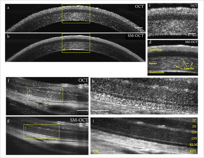
Many approaches have attempted to reduce speckle noise – but the tradeoff is almost always a reduction in image resolution – and they fail to eliminate speckle noise completely in any case. Enter speckle-modulating OCT (SM-OCT) (1). “We wanted to make the speckles dance, so they’d be in a slightly different pattern each time we scanned the tissue,” says Orly Liba, lead author of the study. “And we found a way to do it.” SM-OCT introduces time-varying local phase shifts within the light beam that illuminates the sample, which translates into local phase shifts in the light that’s scattered back from each “scatterer” within each voxel – or “resolution element.” The result? Speckle patterns that change in time and that can be averaged over time to create an image with reduced speckle noise. Crucially, because the sample remains in the same position and because the acquisition is at the same angle with the same set of illumination frequencies, an increase in the number of uncorrelated images does not result in a reduction in resolution – and averaging many images ultimately reduces speckle noise to an undetectable level (relative to other sources of noise, at least).
Adapting existing ophthalmic OCT instruments to become speckle-modulating is actually quite simple. By inserting a diffuser (essentially a plate of glass roughened by randomly etched grooves with a specific size and height) and methodically moving or rotating it between each round of repeated scans, the researchers accessed the optical equivalent of shifting the geographical relationship of the sample’s components a tiny amount for each scan. Modifying the image acquisition software to average successive frames or A-lines produces speckle-free images as seen in Figure 1. What does this all mean? The study’s senior author, Adam De La Zerda, explained, “We showed that you can take effectively any OCT system out there and, with minimal changes, boost its resolution to the point where it can detect anatomical features smaller than the size of a typical cell.”
References
- O Liba et al., “Speckle-modulating optical coherence tomography in living mice and humans”, Nat Commun, 8, 15845 (2017). PMID: 28632205.
