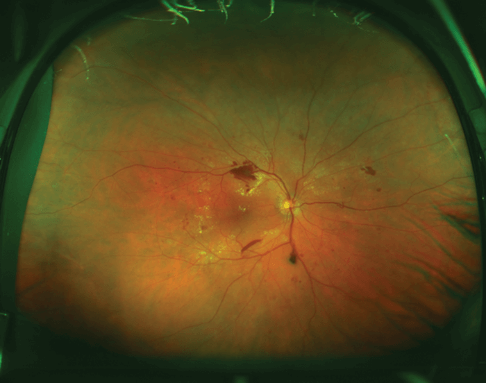
Ultra-widefield (UWF) imaging – which can capture 200° of the retina in a single shot – has been driving advances in the diagnosis and management of retinal disease since its introduction. But what are the new concepts and hot topics in UWF imaging? SriniVas Sadda of Doheny Eye Institute, Los Angeles, CA, USA, shared three new exciting developments. First on the agenda: integrated OCT and Optos UWF imaging, which according to Sadda might be the optimal screening tool or companion for total examination: “Being able to acquire comprehensive information on central and peripheral imaging in one stop is practical – we may be able to perform this on all of our patients.” Next, Sadda shared key consensus recommendations to improve consistency of terminology relating to widefield and UWF imaging, and newer modalities, such as OCT and OCT-A: incorporation of anatomical landmarks within the retina for defining widefield imaging (see Box); improvement of inter-device consistency across various imaging modalities; incorporation of montage and asymmetry into the image description; and introduction of the new term panretinal to represent a 360° ora-to-ora view of the retina. “This proposed system based on anatomical landmarks is more clinically practical and allows us to have consistency when we’re talking about different devices,” explained Sadda.
- Posterior pole – retina within the arcades and just slightly beyond
- Midperiphery – region of retina up to the posterior edge of the vortex vein ampulla
- Far periphery – region of retina anterior to the vortex vein ampulla
- Widefield – single capture image(s) centered on the fovea and capturing retina in all four quadrans posterior to and including the vortex vein ampulla
- Ultra-widefield – single capture view of the retina in the far periphery in all four quadrants
- Panretinal – single capture 360° ora-to-ora view of the retina
Finally, Sadda discussed identifying peripheral non-perfusion as an application of UWF fluorescein angiography (FA) imaging. Many groups have published correlations between ischemic index – the precise area of non-perfusion – and pathology, such as macular edema, and shown that targeted retinal photocoagulation (TRP) may improve outcomes and reduce recurrence rate of anti-VEGF therapy (1)(2)(3). But there is inconsistency in the literature: the DAVE trial (CT.gov, NCT01552408) showed no benefit of TRP addition to ranibizumab therapy in patients with DME; and reading center analysis of UWF FA images (40 eyes of 29 patients) from the DAVE study found no correlation between DME severity and the total amount of non-perfusion or ischemic index (4). Re-analysis of these images by masked graders showed that only non-perfusion with leakage positively correlated with the severity of DME, whereas non-perfusion without leakage was negatively correlated. “We need to re-evaluate these areas of non-perfusion as perhaps targeting one type of non-perfusion could actually lead to better outcomes,” he concluded.
The power of the periphery
Lloyd Paul Aiello, of Harvard Medical School and the Beetham Eye Institute at the Joslin Diabetes Center, Boston, MA, USA, also discussed the need to re-evaluate the information that the periphery holds, and shared potential impact of UWF imaging for diabetic retinopathy (DR).
In 1968, during a discussion on the classification of retinopathy at the Airlie House Symposium, William P. Beetham – Aiello’s grandfather – said, “You can get out very much further toward the periphery (and there is a lot of pathology there) than you can get with your fundus photograph. Is that not true?” But back in 1968 there were technical challenges – going out to the equator meant making a montage of multiple fundus photographs, and took about 12 hours per eye. “Now, 50 years on, a single image can be obtained in a quarter of a second, and this has opened many new possibilities,” said Aiello.
Many retinal physicians are aware of the seven standard fields used to guide DR care (5). “This approach however only covers approximately 30 percent of the retina and doesn’t include the periphery. Optos UWF can image 82 percent of the retinal surface.” commented Aiello. Although previous studies have suggested that ETDRS and UWF imaging are comparable for determining DR severity (6), recent analyses of a large study have demonstrated moderate to substantial agreement within the seven standard fields: in the DRCR.net protocol AA study – an ongoing observational four-year masked evaluation of 766 eyes of 386 patients – DR severity by ETDRS photos and masked UWF images matched exactly in 59 percent of images and were within one-step in 97 percent (7).
But does the additional visualized area provided by UWF imaging add benefit to clinical care? According to Aiello, increasing amounts of research are showing that the periphery might uniquely identify subsets of individuals who are at risk of DR progression. Studies have suggested that presence of predominantly peripheral lesions (PPLs), defined as when more lesions are present in the periphery than posteriorly in at least one field (8), might indicate a higher risk of DR progression. In the ongoing DRCR.net protocol AA study, PPLs were present in 41 percent of eyes at baseline, and were found to suggest an increased disease severity of two or more steps in 11 percent of eyes (7). Aiello also presented further evidence suggesting that PPL may reflect the extent and location of retinal non-perfusion, and are associated with the risk of DR progression. In eyes with no or non-proliferative DR (NPDR) at baseline, the risk of four-year DR progression increased in eyes with PPLs at baseline (n=55) compared with eyes without PPLs at baseline (n=54; p≤0.0322); and the risk of PDR onset was increased 4.2-fold in eyes with PPLs at baseline. In eyes with mild NPDR at baseline, nine percent of eyes with PPL developed PDR at four years, compared with only one percent of eyes without PPL; in eyes with moderate NPDR at baseline, ~60 percent of eyes with PPL at baseline developed PDR at four years, compared with only 15 percent of eyes without PPL.
What does this tell us? That UWF PPL evaluation identifies eyes at higher risk of progression within the same ETDRS severity level than is possible by standard imaging alone. “The implications of this are profound, and suggest that current standard retinopathy grading approaches may require modification to incorporate peripheral evaluation for optimal DR risk assessment,” commented Aiello. Current guidelines from the AAO recommend yearly exams for patients with mild or moderate NPDR with no macular edema, but these guidelines do not include peripheral assessments (9). Furthermore, extended follow-up intervals have been suggested for telemedicine programs – but these do not routinely evaluate the periphery. “Not only do these findings clearly imply that follow-up intervals should not be extended for patients with mild or moderate NPDR without first evaluating the retinal periphery, but there are implications for assessing patient need for treatment and clinical trial designs,” said Aiello.
According to Aiello, the key now is to be aware of the information that the periphery holds – and to be aware of how to consider this information when determining DR severity and clinical follow-up intervals. “We’re fortunate to be working during a time when there’s an explosion in technology. Our ability now to non-invasively and easily assess the retina and the periphery in incredible detail holds great potential in allowing us to better care for our patients in a more comprehensive manner – and to further reduce the loss of vision that’s so closely associated with diabetic eye disease.”

References
- M Singer et al., Retina, 34, 1736–1742 (2014). PMID: 24732695. MM Muqit et al., Acta Ophthalmol, 91, 251–258 (2013). PMID: 22176513. Y Takamura et al., IOVS, 55, 4741–4746 (2014). PMID: 25028357. W Fan et al., Am J Ophthalmol, (2017). PMID: 28579062. Early Treatment Diabetic Retinopathy Study Research Group, Ophthalmology, 98, 823–833 (1991). PMID: 2062515. PS Silva et al., Am J Ophthalmol, 154, 549–559 (2012). PMID: 22626617. DRCR.net, “Protocol AA”, JAMA Ophthalmol, [in press], (2017). PS Silva et al., Ophthalmology, 122, 2465–2472 (2015). PMID: 26350516. AAO Preferred Practice Pattern. “Diabetic retinopathy” (2017).
