To coincide with the announcement of our new Hall of Fame inductees – which sees David Huang, co-inventor of optical coherence tomography (OCT), and Dennis Lam, leading clinician, entrepreneur, and philanthropist, added to the esteemed roll-call – The Ophthalmologist recently convened a KOL roundtable to explore the transformative power and future potential of OCT.
For this special event, Huang and Lam were joined by Ophthalmologist Power Listers (see sidebar), who discussed their observations and experiences of how OCT technology has revolutionized the practice of ophthalmology and patient care.
In part three, we ask the panel, “How do you envision AI enhancing OCT’s capabilities, especially in terms of image analysis, diagnostics, or personalized treatment plans? Could AI potentially make OCT even more valuable in clinical settings?”
The panel
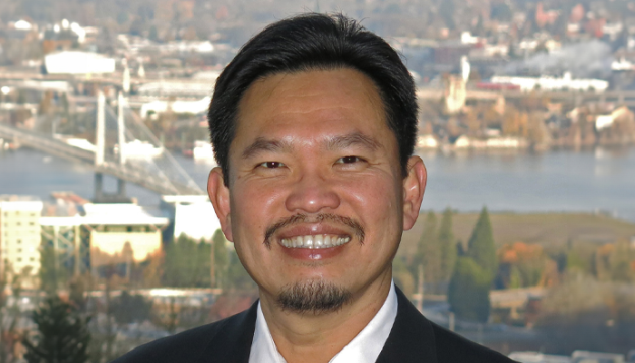
David Huang, Associate Director and Director of Research of Casey Eye Institute, the Wold Family Endowed Chair in Ophthalmic Imaging, Professor of Ophthalmology and Professor of Biomedical Engineering at Oregon Health & Science University (OHSU).
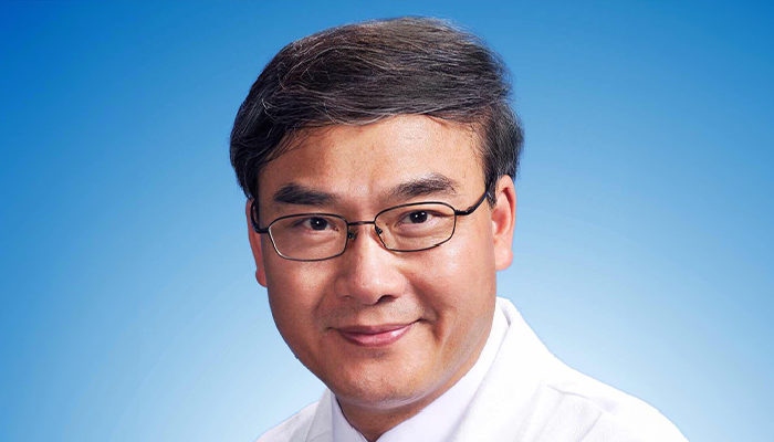
Dennis Lam, President, Hong Kong C-Mer International Eye Care Group; Honorary Director of the Zhongshan Ophthalmic Center (ZOC) of Sun Yat-Sen University, Guangzhou, China; Director of ZOC’s State Key Laboratory; Secretary-General and CEO of APAO and APVRS.
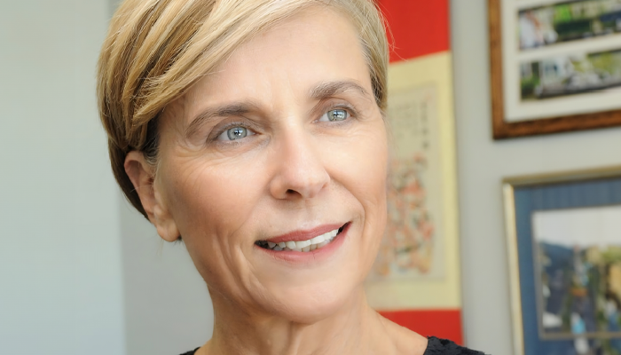
Anat Loewenstein, Chairman of Ophthalmology, Sourasky Medical Center, Vice Dean, Faculty of Medicine, Tel Aviv University; Sidney Fox Chair of Ophthalmology, Tel Aviv University, President of the Israeli Ophthalmological Society.
Andrzej Grzybowski, Professor of Ophthalmology and Head of Institute for Research in Ophthalmology, Foundation for Ophthalmology Development, Poznan, Poland.
Keith Barton, Consultant Ophthalmologist, Moorfields Eye Hospital; Professor of Ophthalmology, UCL; Co-Chair, Ophthalmology Futures Forums, London, UK.
Michael F. Chiang, Director, National Eye Institute, National Institutes of Health, Bethesda, MD, USA.
Nadia K. Waheed, Professor in Ophthalmology, Tufts University School Of Medicine, Boston, Massachusetts, USA.
The panel
David Huang, Associate Director and Director of Research of Casey Eye Institute, the Wold Family Endowed Chair in Ophthalmic Imaging, Professor of Ophthalmology and Professor of Biomedical Engineering at Oregon Health & Science University (OHSU).
Dennis Lam, President, Hong Kong C-Mer International Eye Care Group; Honorary Director of the Zhongshan Ophthalmic Center (ZOC) of Sun Yat-Sen University, Guangzhou, China; Director of ZOC’s State Key Laboratory; Secretary-General and CEO of APAO and APVRS.
Anat Loewenstein, Chairman of Ophthalmology, Sourasky Medical Center, Vice Dean, Faculty of Medicine, Tel Aviv University; Sidney Fox Chair of Ophthalmology, Tel Aviv University, President of the Israeli Ophthalmological Society.
Andrzej Grzybowski, Professor of Ophthalmology and Head of Institute for Research in Ophthalmology, Foundation for Ophthalmology Development, Poznan, Poland.
Keith Barton, Consultant Ophthalmologist, Moorfields Eye Hospital; Professor of Ophthalmology, UCL; Co-Chair, Ophthalmology Futures Forums, London, UK.
Michael F. Chiang, Director, National Eye Institute, National Institutes of Health, Bethesda, MD, USA.
Nadia K. Waheed, Professor in Ophthalmology, Tufts University School Of Medicine, Boston, Massachusetts, USA.
Andrzej Grzybowski: I became fascinated about 15 years ago when I realised that OCT can be used to get an insight not only into the eye, but into the other parts of the body as well. So the eye becomes a window maybe not to the soul, but perhaps to the brain and to the whole body. There’s a growing number of studies showing that with OCT we can follow changes in the retina that are related to neurodegenerative disorders.
It’s now more than 25 years since Vincenzo Parisi published his paper on using OCT to establish that a correlation exists between optic nerve fiber layer thickness and the retina or the retinal pathway function in multiple sclerosis, and since then we have seen OCT also applied to the study of Alzheimer’s disease, Parkinson’s disease, psychiatric disorders such as schizophrenia, and many more conditions. My colleagues and I published a book on OCT and neurodegenerative disorders, the third edition of which is out soon.
Now AI is becoming an important tool in combination with OCT. A few years ago I was part of a study comparing the accuracy of deep learning in detecting glaucoma using optic nerve head fundus photos versus OCT. We found that the performance of the model based on fundus pictures was slightly better than OCT, which was a bit of a surprising result. But with AI, I expect that we will be able to find more information with OCT in the future; we will be able to recognize what is presently unrecognizable. We were able to do this sometimes with the fundus images, which could point to Alzheimer's disease or even the age or sex of the patient. We don't really understand how the AI model made these decisions, but I hope we will be able to gain more information from OCT scans in the future. Of course, the major limitation for AI is access to a good data set. With fundus sometimes it's easier, but it's still a huge limitation with OCT in terms of systemic disorders.
Keith Barton: Yes, thinking about the future my immediate thought was not about how we're collecting the data, but who's making the decisions. AI is talked about a lot these days, but if you listen to what people such as the AI gurus like Geoffrey Hinton and Mustafa Suleyman are saying, it can actually be quite frightening.
Dennis Lam: I think AI is the future and will shape the whole of clinical practice. We talked about ophthalmology in 2040, but in terms of AI, even 2030 is already quite a long time in terms of advancement and development. OCT certainly will have lots of roles to play, particularly regarding optic nerve scanning. For macular images it's already very advanced, but I believe it can go a lot further in optic nerve scanning – for example, for glaucoma in high myopia. It’s quite difficult to make a diagnosis, especially with normal tension glaucoma; it's not just the thickness but the pattern. If we integrate AI with these OCT images, I believe we will be able to make an earlier diagnosis, and the monitoring will be much more accurate.
David Huang: I agree that one of the most exciting frontiers is the use of OCT of the eye to diagnose neurological and cardiovascular diseases under the rubric of “oculomics”. It could be something that's fairly simple. If you look at multiple sclerosis, the use of OCT in neurology is still primitive compared to the way we use it in glaucoma. The official diagnostic criteria for OCT retinal nerve fiber layer measurement in MS involves looking at the left-right eye asymmetry, which is fairly specific but one of the least sensitive ways of detecting loss. If they could simply switch to looking at the temporal nerve fibre layer loss or macular ganglion cell loss, it would be a lot more sensitive. So in this case, some change in methodology would suffice. But I do believe in the long run that AI will be the answer, because family practice doctors don't really have time to learn all the nuances of nerve fibre layer analysis and the macular anatomy, or to understand ophthalmic imaging technology like retina or glaucoma specialists; they need the simple interpretation that AI can provide.
The bottleneck there of course is that we don't have big OCT datasets like we do with fundus photography, and there's a Tower of Babel of different OCT and OCT angiography formats that Michael Chiang at the National Eye Institute is trying to standardize so that development of AI can proceed. I certainly hope that we can make headway there in the near term.
Final thoughts about the future of OCT?
Michael Chiang: We thrive as a field when we do work that matters to eye doctors, obviously, but also that matters to doctors and scientists more broadly. Following the amazing work by David Huang in imaging that applies to many areas of medicine, and the first FDA-approved AI algorithm in any field of medicine for diabetic retinopathy, I think oculomics has very exciting potential. It’s going to be great to think through how we can take advantage of the eye as a “window to the body and a window to expand other areas of science” in the 21st century.
Nadia Waheed: I'm a huge technophile and I believe that if we're able to get through the Tower of Babel of data, standardize all the data that we're getting, and leverage the full force of that to both oculomics as well as to applications within the eye, then we’re on the cusp of breakthroughs, the likes of which we've probably never seen before.
Keith Barton: I think AI will probably apply to glaucoma more than retina, because with glaucoma you've got to tie together the field, the pressure, the corneal thickness, the three different ways of measuring the nerve fibre layer, the Bruch’s membrane opening. So there's a real role for an algorithm to take all that and put it together.
Dennis Lam: AI will certainly be a leading technology and integrating it into eye investigations such as OCT is very important. In terms of systemic diseases, the molecular diagnostic component will also be very important. So I guess the future will be multimodal, integrating a number of different investigations to get to a quick, timely, cost-effective and accurate diagnosis.
Anat Loewenstein: AI is needed to help us with the multiple images that home OCT will create. It's not possible for us to deal with all this data alone. As Keith said, we also need more data relating to visual acuity and the way that it correlates with the fluid and so forth, so there is still a lot to do.
Andrzej Grzybowski: I think we need more collaboration over competition in AI. This has not been the case so much in the last 10 years, but there are now a few more initiatives based on collaboration, and I believe global collaboration will have a real impact.
David Huang: I'm really glad to see all this enthusiasm for OCT and AI, and I agree that we're going to see fantastic advances in these two technologies in the next decade. OCT was propelled by advances in photonics and optical communications in the 1990s and by things like high-speed cameras in the 2000s. I think AI will propel it forward again now. We are going to see different types of OCT applications and see the technology do things in the next 10 years that we could not do before, so it's going to be another exciting decade.
Click on the links for Part 1 and Part 2 of this roundtable discussion.
Join leading ophthalmology experts, including OCT co-inventor David Huang, as they discuss how home OCT will change patient care.
