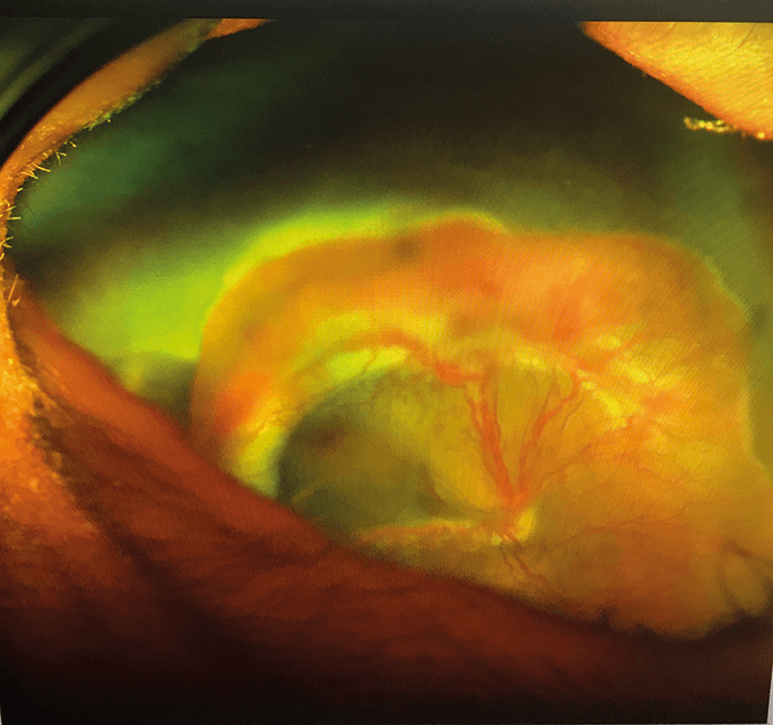
Diffuse Melanoma “This patient has diffuse ocular melanoma of the left eye. Notice the diffuse pigmentation on the bulbar conjunctiva nasally, temporally, and inferiorly.” Zach Dupureur, Ophthalmic Photographer and Research Photographer, and Abu-Bakar Zafar, Ophthalmologist, Carle Foundation Hospital, IL, USA.
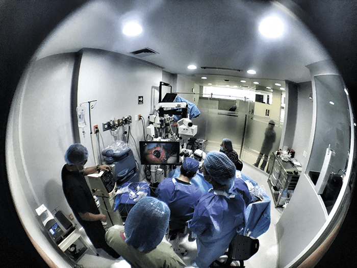
Intrepid Vitrectomy These images document the case of premature baby girl with stage 4a retinopathy of prematurity (ROP) who underwent vitrectomy – the first vitrectomy ever performed on such a young baby in Mexico using heads-up surgery technology. “The surgery was challenging, not only technically, but because of the logistics it involved; no ophthalmology hospital had a neonatal intensive care unit (NICU) and no NICU had equipment for eye surgery.” Maria Ana Martinez-Castellanos, Pediatric Retina Surgeon, Asociación para Evitar la Ceguera en Mexico, Mexico City, Mexico.


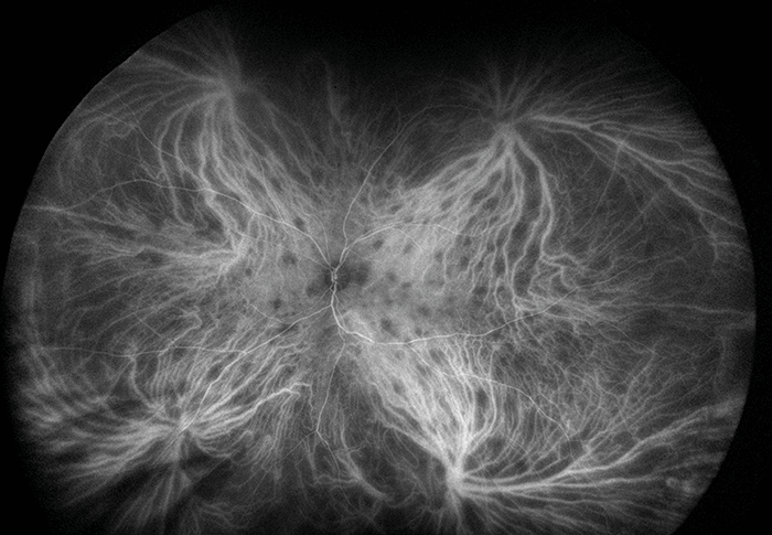
Birdshot Choroiditis Ultra-widefield indocyanine green angiography image of birdshot choroiditis demonstrating lesions in the central and peripheral retina. George Ko and Rahul Mandiga, Retina Insitute of Washington, Renton, WA, USA.

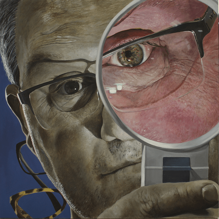
Stephen with a Magnifier
This painting (acrylic on canvas, 2014) is part of Lucy Burscough’s “Look200” series which explores color vision deficiency. The subject is Stephen Golding, a dispensing optician at the UK’s Manchester Royal Eye Hospital. Standard vision color range is shown within his magnifier, while outside is a simulation of the colors seen by someone with severe deuteranopia. The image was developed using Kazunori Asada’s Chromatic Vision Simulator app and was painted in the waiting areas of clinics at Manchester Royal Eye Hospital as part of an Arts Council England funded “arts for health” residency. The whole series and Lucy’s other vision related artwork can be seen at www.LucysArt.co.uk.
Lucy Burscough, Artist, Manchester, UK.

