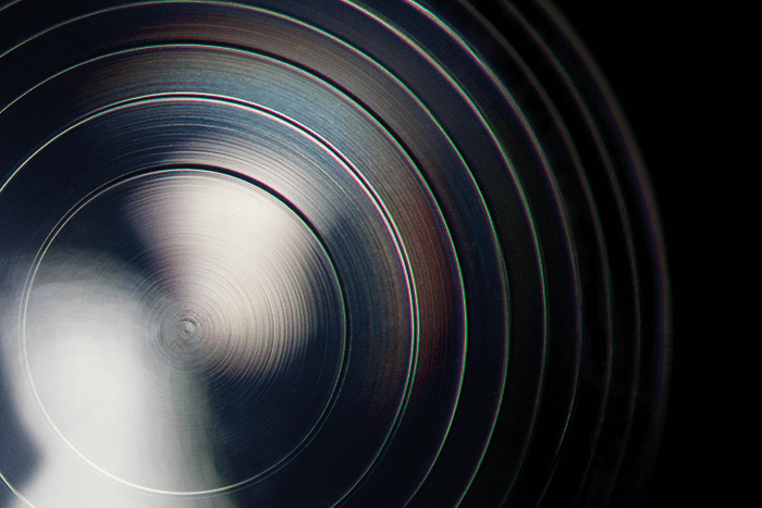
Focusing on Multifocality Image of a extended depth of focus IOL, taken after YAG capsulotomy. Karl Brasse, Ophthalmologist, Vreden and Matthias Gerl, Augenklinik Ahaus, Germany.
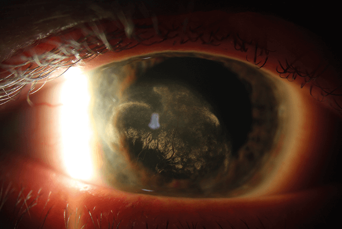
Red and Ruptured
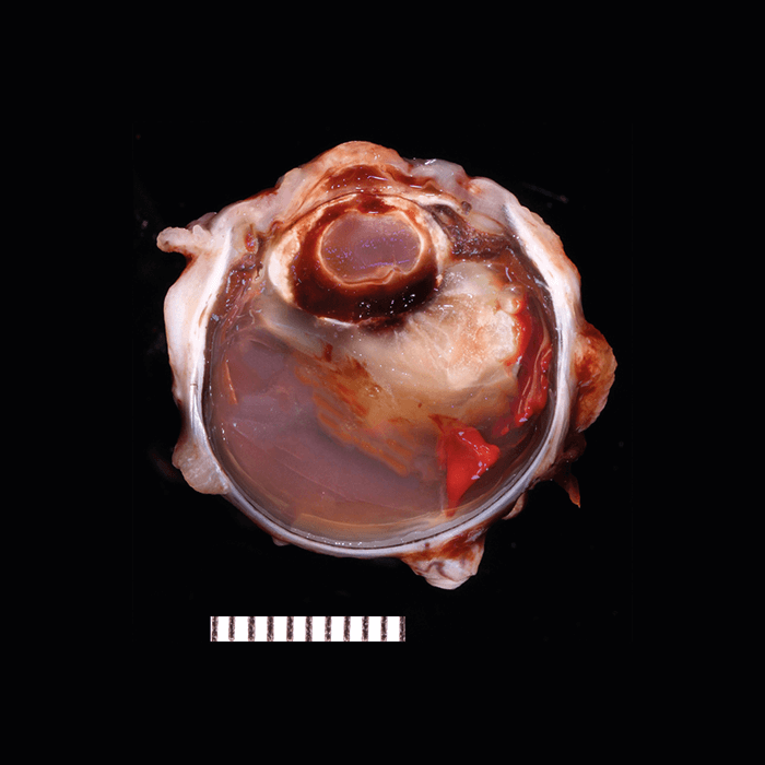
Canine globe with chronic intralenticular hemorrhage, evidenced grossly as red discoloration within the lens capsule. Histologically, blood vessels extended into the lens and percolated through the hemorrhage. The lens capsule was also ruptured and there was a granulomatous phakitis. The blood vessels extending from the optic nerve head remain unidentified. A diagnosis of persistent fetal vasculature (persistent hyperplastic tunica vasculosa lentis and/or persistent hyperplastic primary vitreous) was made.
The Comparative Ocular Pathology Laboratory of Wisconsin (COPLOW), USA. Further images can be found on the COPLOW Facebook page.
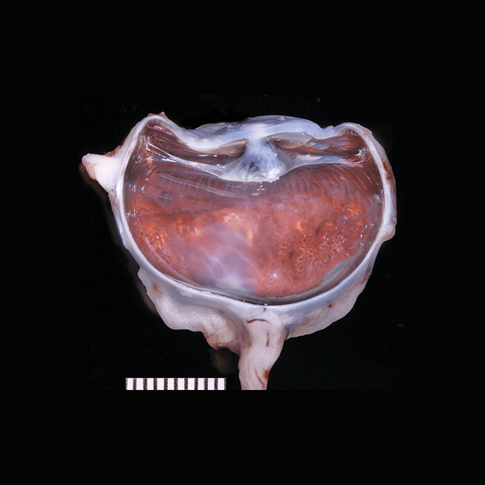
Chamber Cleavage Anterior segment abnormalities can be seen in this canine globe with anterior chamber cleavage syndrome. The small, malformed lens blends with the posterior cornea. Additionally, the iridocorneal angle structures (ciliary cleft and trabecular meshwork) were malformed with no aqueous humor outflow tracts, which was termed anterior segment dysgenesis. The Comparative Ocular Pathology Laboratory of Wisconsin (COPLOW), USA. Further images can be found on the COPLOW Facebook page.
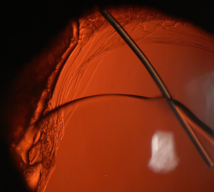
Coastal Plains
A bright beam of light and sclerotic scatter are used to expose corneal irregularities (above).
Houston Sharpe III, Ophthalmic Imaging Specialist, UNC Kittner Eye Center, Chapel Hill, NC, US
Lyon & Healy Retroillumination of a displaced IOL. Houston Sharpe III, Ophthalmic Imaging Specialist, UNC Kittner Eye Center, Chapel Hill, NC, USA.
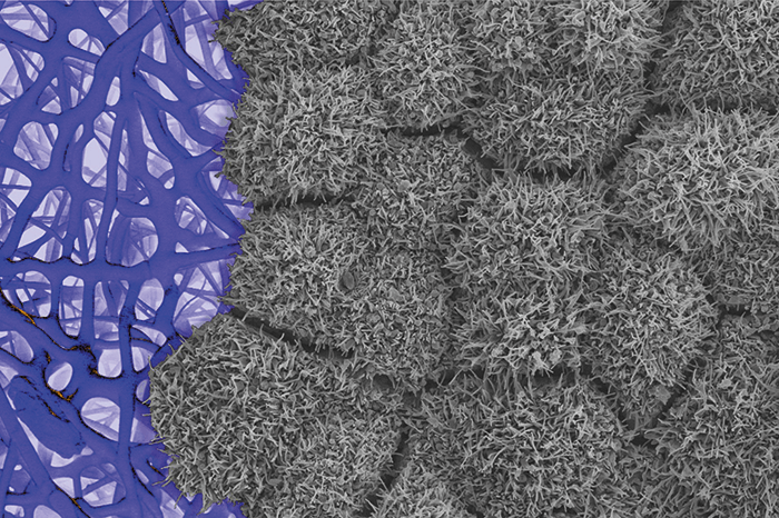
RPE SEM Scanning electron micrograph of induced pluripotent stem-cell-derived RPE cells growing on a nanofiber scaffold. Kapil Bharti, National Eye Institute/NIH.
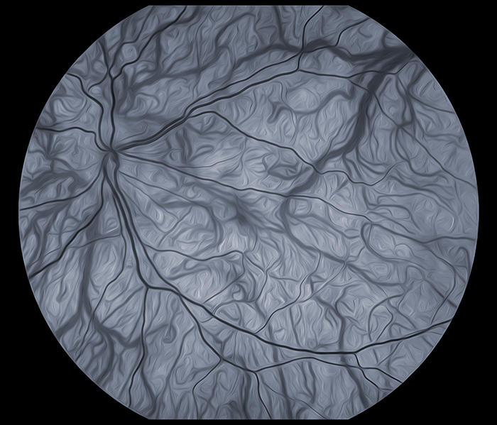
Greyscale Fundus Fundus photo of retina in person with ocular albinism (top right). Kapil Bharti, National Eye Institute/NIH.
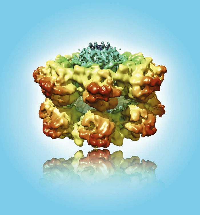
Revealing Retinoschisin An essential adhesion protein in the retina, the loss of retinoschisin results in a form of macular degeneration in young males called X-linked retinoschisis. Bernard Heymann, NIH.
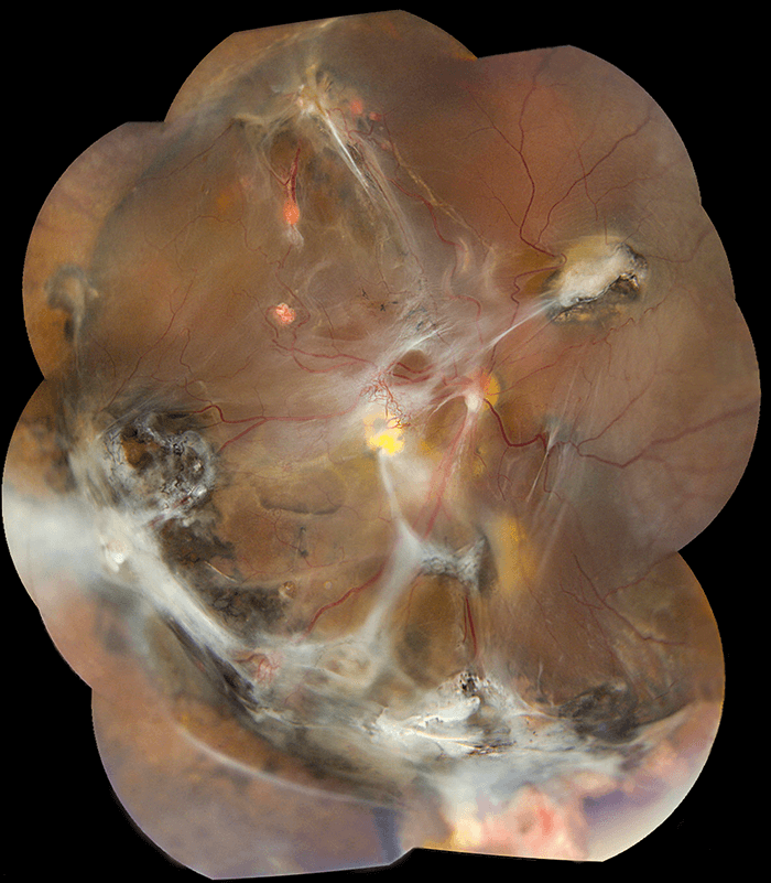
All Together Now Montage photography of Von-Hippel Lindau Syndrome. Mike Arango, National Eye Institute/NIH.
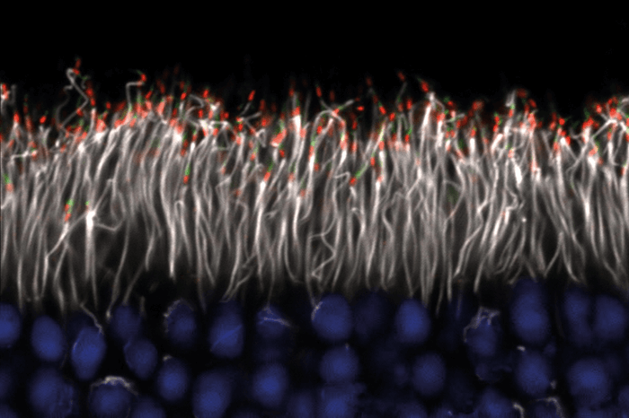
Cilia and Cilia The cellular structure in photoreceptors that capture light is derived from a modified cilium. Fluorescent labeling for RPGR, a protein commonly mutated in retinitis pigmentosa, shows the photoreceptor cilia (red) within the mouse retina. Additional ciliary structures (green), the ciliary rootlet (white), and nuclei (blue) are also visualized. Jessica Gumerson, National Eye Institute/NIH.