Globally, cataracts are responsible for just over half of all cases of blindness and cause severe disability (1). In the US, more than half of all people aged 80 years or older will have a cataract (2) – and treatment costs in 2004 were estimated to be $6.8 billion (3).
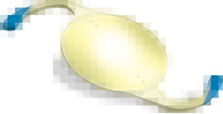
Having said all that, today’s cataract surgery is normally straightforward to perform, and restores good vision almost immediately. Cataract surgery also provides an opportunity for surgeons to correct astigmatism at the same time as removing cataracts, by replacing the crystalline lens with a toric intraocular lens (IOL). However, for toric IOLs to work optimally, they require rotation to be on-axis with the patient’s astigmatism – even slight misalignments can have big impacts upon refractive outcomes (4). Here’s what I do to ensure that my patients receive the best possible refractive outcomes after surgery.
Tip 1: X Marks The Axis
On the operating table, mark the axis of alignment either before surgery or after removal of cataract and stabilization of the chamber. This is important if your view of the lens markings is impeded by pupillary constriction during the procedure or exuberant corneal hydration when sealing the section at the end of the procedure.
Tip 2: Variance Is A Red Flag
Beware marked differences in k values between a patient’s eyes unless there is an obvious cause such as amblyopia or corneal pathology. Repeat if necessary.
Tip 3: Avoid Anesthesia
Do not instill local anesthetic into to the eye prior to performing k readings. This disrupts the corneal epithelium and causes the cornea to dry out – a source of error in k readings. Don’t perform applanation tonometry either, another potential source of k value errors. It is the quality of the tear film that ensures accuracy.
Tip 4: Get Your Bearings
Mark the three and nine o’clock positions of eye with a fine tipped marker, with the patient sitting up to eliminate cyclotorsion.
Tip 5: Look To The Light
Make sure the patient is looking at the operating microscope light to remove the effects of parallax.
Tip 6: Read, And Read Again
To ensure precision for lens calculation, obtain several readings. Repeat on different days if measurements are variable; this is especially important in the presence of high degrees of corneal astigmatism.
Tip 7: Incisive Incisions
When making the corneal incision, ensure accurate placement in the normal corneal meridian based on your audited results.
Tip 8: Soft Shell Shuffle
In the Arshinoff Soft Shell technique, you partially fill the bag with OVD whilst completely filling the rest of the chamber prior to lens insertion. Fill the remainder of the bag with balanced salt solution (BSS) beneath the OVD, then inject the IOL. With this technique virtually all the OVD is anterior to the IOL and removal is rapid and complete with I/A.
Tip 9: Successful Completion
Following removal of OVD ensure that the IOL is in the correct axis. Redial into correct position if necessary. Next, ensure adequate chamber stabilization with stromal hydration of the corneal section. It is the combination of complete OVD removal and chamber stabilization that ensures rotational stability of the IOL. Remember not to mask the lens markings with your stromal hydration when sealing the section. Finally, inject intracameral cefuroxime via the paracentesis and make a final check that the lens is ‘on axis’.
Tip 10: OVD: Out, Out, Out!
To ensure complete removal of the ophthalmic viscosurgical device (OVD), drop the bottle and use a low vacuum when the tip of the irrigator/aspirator (I/A) is behind the IOL.
David is a consultant ophthalmic surgeon & retina specialist, Aut Even Hospital, Kilkenny, Ireland & Barringtons Hospital, Limerick, Ireland, a researcher at the University of Liverpool, UK, and a visiting professor at Florida University.
References
- World Health Organization, “Prevention of Blindness and Visual Impairment: Priority Eye Diseases”. ; accessed December 16, 2013.
- Prevent Blindness America/ National Eye Institute, “Vision Problems in the U.S.: Prevalence of Adult Vision Impairment and Age-Related Eye Disease in America” (2008). ; accessed December 16, 2013.
- Rein, P Zhang, KE Wirth et al., “The economic burden of major adult visual disorders in the United States.” Arch. Ophthalmol, 124(12), 1754-60 (2006).
- H Jin, IJ Limberger, A Ehmer, et al., "Impact of axis misalignment of toric intraocular lenses on refractive outcomes after cataract surgery", J Cataract Refract Surg. 2010, 36(12), 2061-72 (2010).
