Conexiant
Login
  • Corneal Physician
  • Glaucoma Physician
  • New Retinal Physician
  • Ophthalmology Management
  • Ophthalmic Professional
  • Presbyopia Physician
  • Retinal Physician
The Ophthalmologist
  • Explore

    Explore

    • Latest
    • Insights
    • Case Studies
    • Opinion & Personal Narratives
    • Research & Innovations
    • Product Profiles

    Featured Topics

    • Anterior Segment
    • Glaucoma
    • Retina

    Issues

    • Latest Issue
    • Archive
  • Subspecialties
    • Cataract
    • Cornea
    • Glaucoma
    • Neuro-ophthalmology
    • Oculoplastics
    • Pediatric
    • Retina
  • Business

    Business & Profession

    • Professional Development
    • Business and Entrepreneurship
    • Practice Management
    • Health Economics & Policy
  • Training & Education

    Career Development

    • Professional Development
    • Career Pathways

    Events

    • Webinars
    • Live Events
  • Events
    • Live Events
    • Webinars
  • Community

    People & Profiles

    • Power List
    • Voices in the Community
    • Authors & Contributors
  • Multimedia
    • Video
    • Podcasts
Subscribe
Subscribe

False

Advertisement
The Ophthalmologist / Issues / 2015 / Jun / Target Practice
Retina Glaucoma

Target Practice

A light box and a 35 mm slide could be all you need for therapeutic laser target practice

By Michael Schubert 6/8/2015 1 min read

Share

Figure 1. 35mm retinal slide on a lightbox.

How do you train an ophthalmologist to use a laser for the first time? If you don’t have access to a computer-based simulator, there are some imaginative alternatives. Take laser photocoagulation on a “simulated fundus” as an example (1) – a piece of paper that has been photocopied black on one side, and some basic retinal vasculature drawn on in pen on the white side, is taped to the laser headpiece, and suffices as an extremely inexpensive practice medium – it’s okay, but it’s certainly something that can be improved upon.

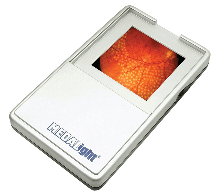
That’s exactly what UK-based ophthalmologist Seema Arora and Leadership Fellow in Simulation, Polly Dickerson, decided to do (2). Instead of attaching paper to a retinal laser’s headpiece, they attached 35 mm photographic slides depicting retinal pathologies and a small battery-operated lightbox (Figure 1) to the laser machine’s forehead rest. How does it work? Trainees apply the laser beam directly to the slides without the need for a contact lens; they can try out different pulse durations and investigate treatment options for various procedures. Images can be projected to multiple viewers before and after treatment, which supports group training and discussion – and Arora and Dickerson have developed a “recipe book” of faculty requirements and debriefing discussions in their training materials to maximize the benefits of the slides. Slides can also be printed with photographs of the angle structure – ideal for practicing selective laser trabeculoplasty. Although the 35 mm slides are both inexpensive and easy to use, there is one big drawback; laser burn intensity cannot be titrated on them. Nevertheless, Arora and Dickerson view the slides as “a cost-effective and widely available tool that has the potential to democratize access to simulated laser training.”

References

  1. AW Cohen, et al., “Learning to use the PASCAL laser system; a part of on-going professional development” (2009). Available at: http://bit.ly/1PLdiNh. Accessed May 26, 2015.
  2. S Arora, P Dickerson, “35 mm slides – a novel target to simulate laser treatment”. Poster presented at the Royal College of Ophthalmologists Annual Congress; May 19–21, 2015; Liverpool, United Kingdom. Available at: http://bit.ly/1Ey1rpQ. Accessed May 26, 2015.

About the Author(s)

Michael Schubert

While obtaining degrees in biology from the University of Alberta and biochemistry from Penn State College of Medicine, I worked as a freelance science and medical writer. I was able to hone my skills in research, presentation and scientific writing by assembling grants and journal articles, speaking at international conferences, and consulting on topics ranging from medical education to comic book science. As much as I’ve enjoyed designing new bacteria and plausible superheroes, though, I’m more pleased than ever to be at Texere, using my writing and editing skills to create great content for a professional audience.

More Articles by Michael Schubert

Related Content

Newsletters

Receive the latest Ophthalmology news, personalities, education, and career development – weekly to your inbox.

Newsletter Signup Image

False

Advertisement

False

Advertisement

Explore More in Ophthalmology

Dive deeper into the world of Ophthalmology. Explore the latest articles, case studies, expert insights, and groundbreaking research.

False

Advertisement
The Ophthalmologist
Subscribe

About

  • About Us
  • Work at Conexiant Europe
  • Terms and Conditions
  • Privacy Policy
  • Advertise With Us
  • Contact Us

Copyright © 2025 Texere Publishing Limited (trading as Conexiant), with registered number 08113419 whose registered office is at Booths No. 1, Booths Park, Chelford Road, Knutsford, England, WA16 8GS.

Disclaimer

The Ophthalmologist website is intended solely for the eyes of healthcare professionals. Please confirm below: