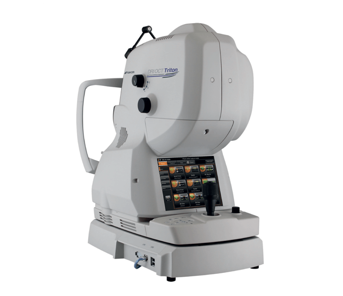
Much of contemporary medicine today depends on the use of medical equipment with advanced technologies to ensure optimal patient outcomes and improved clinical workflows – and ophthalmology is certainly no different. Optical Coherence Tomography (OCT) imaging is widely known as a vital tool for the early detection, prevention, and management of a wide range of eye diseases. OCT technology allows clinicians to quickly and efficiently assess a patient’s eye health so that appropriate treatment can be administered, and, if necessary, to save patients’ vision. Yet, the clinical needs of ophthalmologists in terms of imaging equipment are often complex. Topcon’s TritonTM OCT/fundus camera offers an effective solution, combining both multimodal fundus imaging with the world’s first Swept-Source OCT.
To learn more about how the Triton can benefit an ophthalmologists’ practice, including the improvements it offers over spectral domain OCTs, we talked with Dr. Brett Bielory, ophthalmologist and Medical Director at Riverside Medical Group in New Jersey, about his experience with the Triton.
Dr. Bielory, how has the Triton OCT proven valuable in your practice?
The Triton offers some unique abilities with its Swept-Source technology because it gives a complete picture of the eye from the front to the back. From the anterior segment to the fundus, and with OCT imaging of the retina and optic nerve, you are guaranteed a complete clinical picture of the patient’s eye before they even step into the room. This device is a must-have in the comprehensive clinician’s setting.
What has surprised you about the Triton?
The biggest surprise was how quick, efficient and easy to use the technology is – especially for imaging technicians. It didn’t take long at all for the technicians in my practice to adjust to the Triton. We had a single training session in the morning, and they were “off to the races” in the afternoon, directing patient care.
How is the Triton OCT and fundus camera useful in the clinical assessment and management of your patients?
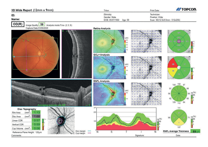
The Triton OCT is capable of generating true-color fundus photos and high-quality OCT scans in a single acquisition. With this capability and its 12mm x 9mm widefield scan function, the Triton offers an excellent ocular health “snapshot” of the posterior globe.
How does the speed of image acquisition compare to other OCT systems you have used?
It is far superior! The Triton OCT uses Swept-Source technology, offering increased scanning speed and allowing for high-quality imaging into the deepest layers of the eye – even through cataracts, hemorrhages, and other complex opacities.
How has the Triton’s swept-source technology impacted your clinical workflow and decision-making ability?
As a clinician, I want to be confident that I am able to diagnose and treat my patients in a comprehensive and efficient manner. The Triton offers me the reassurance of a reliable scan irrespective of ocular pathology. I have found that dense lens opacities and vitreous hemorrhages will not prevent successful imaging of the posterior pole.
Could you describe a case in which the Triton was able to scan an eye you assumed you would not be able to image?
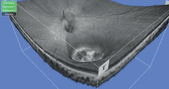
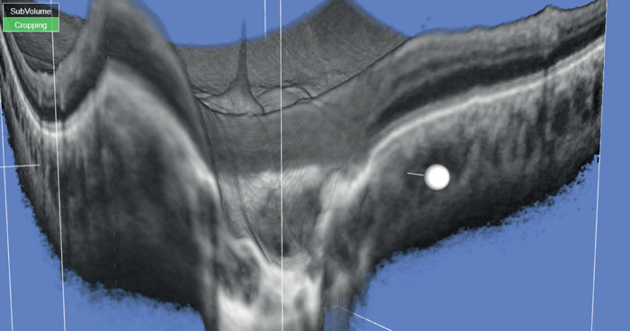
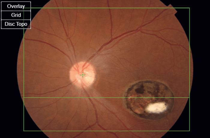
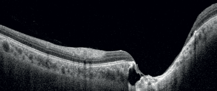
We had a 19-year-old patient with congenital toxoplasmosis and concomitant bilateral horizontal nystagmus. This is a deeply challenging presentation but, using the Triton, we were able to capture a quick snapshot of the posterior pole. Although not perfect, the photos captured were detailed enough to safely document the size of the central macular opacity and determine whether any vitreous opacities were present. See Figures 1A-3.
What is your message to other ophthalmologists who may be looking to purchase an OCT for their practice?
I would go so far as to say that, without one, you are providing substandard service to your patients. In today’s medical world, reimbursements continue to plummet and third-party payers push for doctors to see more patients. We are required to improve documentation and Triton’s images, in combination with Topcon’s Harmony® software, allow you to meet compliance standards. One must incorporate OCT into day-to-day practice to prevent the potential legal liability of missing pathology.
What has been your impression of the Triton’s performance and reliability?
It has been second to none! In my opinion, there is really no reason to consider any other device given the speed and accuracy of each captured image. My patients are impressed with the technology. Clinical throughput is efficient and timely, and I’m able to show my patients any relevant findings using the Harmony software. Patients appreciate the extra moments to review testing completed in the office – and the Triton offers excellent progression analysis and clinical information for providers to interpret and review with patients.
Topcon is very receptive to customer feedback and the needs of clinicians in case of technological difficulties or repairs. We are blessed with excellent sales and technical representatives in our region, which is likely an indicator of the service at a national level, too.
Could you envision both the Triton and its cousin, the Topcon Maestro 2 OCT, playing a valuable role in your ophthalmology practice?
Yes, this combination could be very useful! With higher-volume practices, the Maestro2 OCT adds an element of ease and flexibility with a one-button “touch and shoot” ability to increase efficiency and throughput. The robotic Maestro2 is almost akin to the “set it and forget it” turkey cooker sold in the info-commercial – without heating up the patient, of course!
How would you summarize the key benefits of the Triton OCT?
If I had to sum up in just a couple of words it would be versatility and efficiency – it’s invaluably useful in a whole host of different clinical situations and the speed of image capturing really does make it stand out. When that is combined with the detailed image quality as well as how easily it copes with complex cases, the Triton OCT really is a must-have piece of equipment.
The opinions expressed in this article are the author’s own and do not constitute advice from Topcon on how to diagnose.
MCA5044