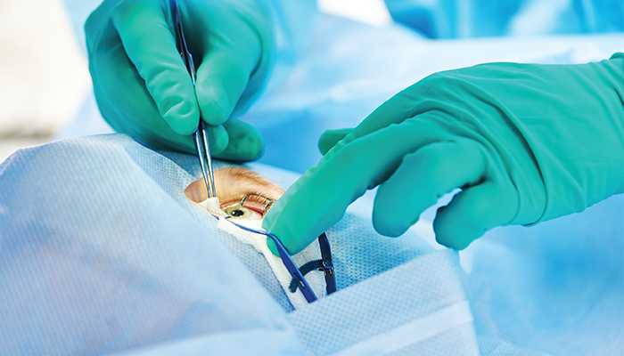
Retinal surgery has gradually become vitreoretinal (VR) surgery; it’s extremely rare today that a posterior segment surgeon performs scleral buckling only. This two-part series, therefore, discusses a handful of highly selected surgical tips related to vitreoretinal procedures. In Part 1, we cover the preparation needed before surgery begins.
1. Counseling
Consultation with the patient is an integral part of any physician-patient relationship – and in VR surgery it is essential. Having the patient sign an “informed consent” form may suffice for legal, but not for medical purposes. The essence of counseling is to present different options to patients and their families, supporting them in making decisions. Paternalistic medicine (“the doctor knows best and will tell you what treatment option to choose”) has no place in contemporary practice.
The conversation between doctor and patient should cover a host of issues, including (but not limited to):
- whether to perform surgery at all
- the patient’s expectations and if they are realistic
- the goals of surgery and the definition of success (what can be gained?)
- the possible complications of the surgery (what can be lost?)
- when to do the surgery
- what kind of surgery
- anesthesia (local versus general)
- the postoperative course (positioning, complications, reoperations etc.)
Each of these items should be discussed in detail, but here we will focus on anesthesia because it is not as straightforward as it may seem.
Both forms of anesthesia have advantages and disadvantages. The advantage of a local anesthesia that is rarely mentioned in textbooks is that the surgeon is able to consult the patient during the operation if an unexpected issue emerges. For example, a surgeon may be detaching the posterior hyaloid in an eye undergoing surgery for a macular pucker when the retina unexpectedly tears at multiple locations. The surgeon completes the operation but feels the need for a longer-term tamponade, which can be a long-acting gas or silicone oil. For the patient, this either means a long period of no vision, then a long period of low vision (through the gas bubble), or a temporary and relatively small refractive change, plus the need for a second surgery (oil removal). Some patients prefer the first option; others prefer the second. Of course, a patient that is awake can make the choice; those under general anesthesia have no option but to accept the consequences of the surgeon’s decision.
The surgeon and the staff
Though the ultimate decision-maker is the surgeon, no surgeon can work alone. The supporting staff includes many people outside the operating room (OR), pre- and postoperatively. But inside the OR there are two key personnel who can greatly influence the success of the operation – the circulator and the nurse.
The circulator must make sure that all instruments and tools can be immediately located when needed, and that these items and their attachments are in a usable condition. One example is the cryo machine – this is rarely needed today, but there are situations where it may be urgently required to treat the retinal periphery or retrieve a nonmagnetic intraocular foreign body of unique size, shape, or surface characteristics.
As for the nurse? “No surgeon is better than his nurse,” so the saying goes. The surgeon must concentrate on a single issue during the operation: the eye. If the surgeon has to struggle with an untrained or inattentive nurse, who repeatedly hands over the wrong instrument or an instrument that needs adjusting, it forces the surgeon to deviate attention from the eye, leading to frustration and an increased risk of intraoperative iatrogenic complications. Conversely, a good nurse not only helps the surgeon to focus solely on the operation, but can also make suggestions that may improve the procedure.
It is crucial, then, for the surgeon to work with a nurse with whom they have an excellent working relationship. With the exception of some unforeseen event, the nurse must remain on hand until the completion of the operation.
The surgical set-up
This fundamental step is too often neglected; most surgeons simply sit down at the microscope and adapt to the previous surgeon’s “leftover” settings. This is wrong for many reasons, so we recommend the steps below.
a) Pedals
Use your dominant foot to drive the microscope pedal. Even if the zoom and focus functions are relatively rarely used, the X-Y joystick is constantly in use. Driving the vitrectomy pedal requires a lot less attention (fewer variables) and can be operated by the non-dominant foot.
b) Posture
Never try to adopt your position to the height of the microscope. First, sit comfortably in the chair with your back straight but not overstretched, your lower legs at ~90 degrees to your thigh, and your feet positioned to easily reach the pedals. Next, adjust the microscope’s height to your position. Then, adjust the height of the operating table to the height of the microscope.
c) Vitrectomy pedal
Many surgeons press down the pedal to activate the aspiration, then they turn their foot outwards to initiate cutting. This puts the foot in an unnatural position, straining the leg muscles. The surgeon must decide whether this is acceptable or choose a different set-up.
d) Hand support
Is it possible to do fine VR surgery without hand support? Yes. But there is no need to overcome unnecessary adversity for the sake of our ego. Hand – and especially wrist – support allows the surgeon to minimize tremor and strain, and focus on the eye and the eye alone.
References
- M Bouaziz, “Shared Decision Making in Ophthalmology: A Scoping Review,” Am J Ophthalmol., 20, 146 (2021). PMID: 34942109.
- F Kuhn, A Grzybowski, “The Role of Counseling in Ophthalmology,” Am J Ophthalmol., 244, vii (2022). PMID: 35568250.
