Figure above: Image courtesy of José Maria Ruiz-Moreno, MD, PhD, FEBO, Head of Ophthalmology, Puerta de Hierro University Hospital, Majadahonda (Madrid), Professor at Medicine School at the University of Castilla-La Mancha (Albacete) in Spain.
Topcon Healthcare (Tokyo, Japan) is the leading manufacturer of Optical Coherence Tomography (OCT) technology (1) and maintains this leadership position in the competitive field of OCT instrumentation. But how has the company achieved this? Topcon’s innovative OCT technology encompasses both spectral domain (SD) and swept-source (SS) OCT applications, which enhances the company’s integration within the market, meeting the needs of both primary and secondary care clinicians.
The technology
Today, it is hard to imagine ophthalmology without OCT. Its rapid, non-contact, in vivo imaging technology is key to diagnostic evaluation and management of disease in both the posterior and anterior segments. The clear benefits of OCT have resulted in a highly competitive marketplace comprised of a number of OCT manufacturers and an expanding range of instruments and features. Initial devices, based on time domain (TD) technology, are increasingly being replaced by second-generation instruments using Fourier domain (FD) technology. FD-OCT has faster scanning speeds, making them less vulnerable to motion artifacts and higher sensitivities. A higher signal-to-noise ratio permits better visualization of low-contrast structures. FD-OCT incorporates spectral domain (SD) technology, which uses a near-infrared superluminescent diode light source and detects signals using a spectrometer. Swept-source (SS) is the leading-edge OCT technology that relies on a swept light source and a single photodiode detector. How can eye care providers make sense of this complexity and choose the best instrument for their needs?
The numbers don’t lie
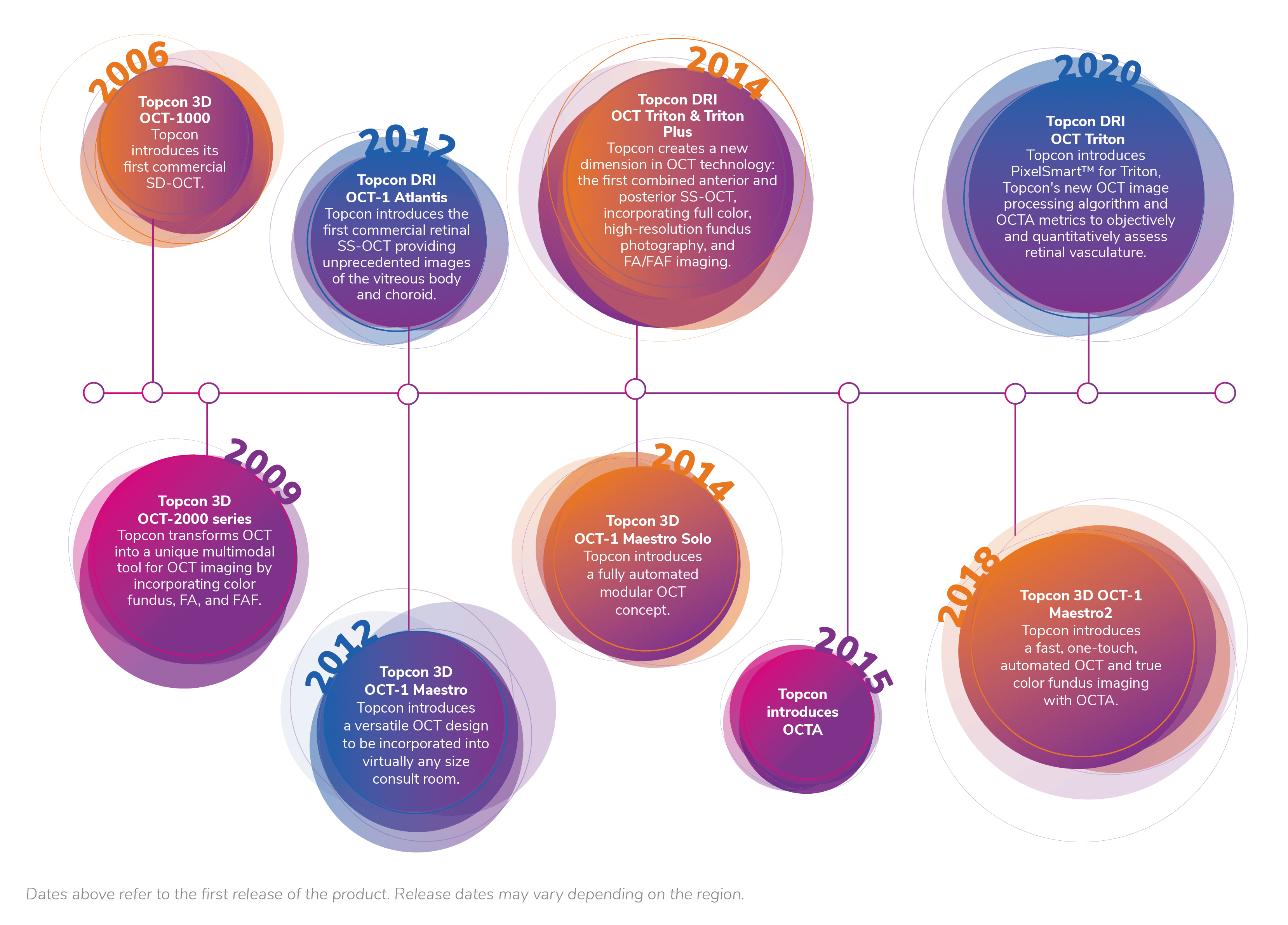
Independent market analysis shows that the OCT instrumentation market leader today is Topcon Healthcare (see Table 1). This has not happened by accident, Topcon Healthcare has continuously invested in technological development (see the timeline). Fumio Ohue, Managing Executive Officer, Global Head of Eyecare Business at Topcon, states, “Since we launched the first SD-OCT in 2006, we have consistently innovated to ensure we meet the growing needs of clinicians across eye care and have been humbled by a response that rapidly took us to the position of market leader.” Ohue asserts, “Topcon will continue to offer cutting-edge technology and innovations to address the full needs of eye care professionals.”
Table 1. OCT instrumentation market share. Topcon Healthcare has higher sales than any other manufacturer and has captured nearly 30 percent of the market (1).
Broad choice, deep expertise, high speed
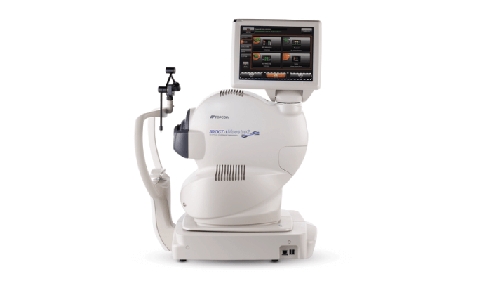
In the SD-OCT field, Topcon’s Maestro2 instrument (see Figure 1) uniquely combines OCT capabilities with a high-resolution, color imaging, non-mydriatic retinal camera. Maestro2 offers fully automated alignment, focus and capture capabilities. Additional features include accelerated workflow, enable image capture, and provide exceptional convenience of operation. Other capabilities include 12 x 9 mm scanning and seven-layer automated segmentation, including the choroid. These features enable measurement and topographical mapping of the optic nerve and macula in one scan. The device also has a “compare” function to allow serial monitoring of OCT images. Finally, the Maestro2’s space-saving footprint makes it compatible with even the smallest office setting.
Jacqueline Sousa Asam, MD, Medical Science Liaison and Clinical Trainer at Topcon Healthcare, comments on the company’s technology, “Commercially available SD-OCT devices use a relatively short wavelength, allowing for good axial resolution. Topcon Healthcare combines this technology with a fully automated acquisition mechanism in the Maestro2. In comparison, commercially available SS-OCT devices take advantage of longer wavelengths and higher-frequency signal detection capability, resulting in larger image depth and less sensitivity roll-off due to the principle of Swept Source that can decompose high frequencies and lower scattering loss. Topcon DRI OCT-1 Atlantis was the first SS-OCT in the market and combined new cutting-edge technology with high speed, providing high-quality images within a short acquisition time. In scientific studies, SS-OCT showed better choroidal visualization than SD-OCT (1, 2).”
We have always been early adopters of new technology at Taylor Biddle Opticians, and had our first Topcon OCT in 2009. We have upgraded it a couple of times in the interim, but recently decided to try the Maestro2. At first, we were reluctant to let go of the joy-stick, but now we are really confident and precise in the capture. It is very quick and efficient. The image quality is fabulous, even through a small pupil or opacities. It is so much better when compared with the same patient from the previous year on the old machine. There are additional features such as the widefield scan that captures both the disc and macula in one scan and the Hood report which gives so much data for glaucoma analysis.
Tracey Taylor, BSc (Hons) MCOptom Optometrist, Director of Taylor Biddle Opticians Ltd, UK
“I use SS-OCT in my daily practice because its longer wavelength allows me to better visualize the layers of the back of the eye, from the vitreous to the choroid, even, through some media opacities.”
Luis Arias, MD, PhD, Head of Retina Department of University Hospital of Bellvitge and Aggregate Professor of Ophthalmology, University of Barcelona, Spain

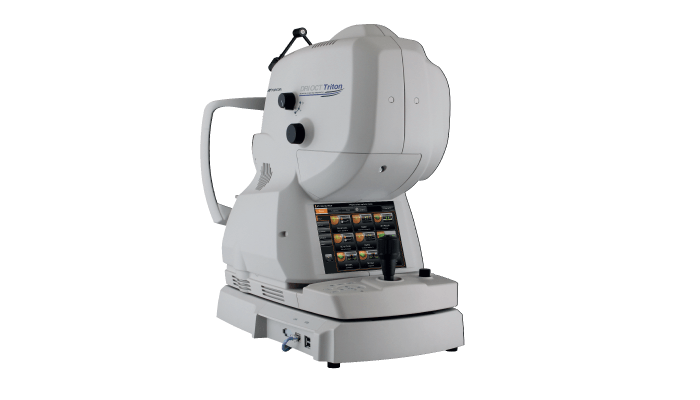
DRI OCT TritonTM series (see Figure 2) offers a truly unique combination of capabilities. It uses optimized, long-wavelength light, 1,050 mn, which provides deep scanning capabilities sufficient to penetrate media opacities, such as cataracts and haemorrhages. At the same time, it comes with the next-generation SMARTTrackTM eye tracking system; this, combined with ultra-fast SS technology (at 100,000 A-scans/s), maximizes data acquisition. By providing more B scans image within a 3D data cube, Triton supports better clinical decisions. Importantly, the scanning line is virtually invisible to patients, thus assisting fixation and reducing movement artifacts, making Triton a natural choice also for younger or geriatric patients. From a physician’s perspective, Triton offers extraordinary simplicity and convenience by capturing comprehensive retinal and choroidal data in a single scan (the system automatically detects seven boundaries, including the chorioscleral interface).
Triton’s unique “5-in-1” diagnostic tool provides outstanding real-world utility for FA, FAF, OCTA, importation of indocyanine green angiography (ICGA) and capture of high-quality, true-color fundus images. Triton also captures true-color fundus images in 3D using stereo photography and provision of wide coverage of the retina from the central macular area to the periphery (almost the complete fundus) by virtue of the automosaic panorama facility.
“The Triton was the first SS-OCT capable of deep-range imaging and still exceeds imaging available from spectral domain devices. It can image from the vitreous to the choroid in a single scan.”
Richard Spaide, MD, Retina and Vitreous Specialist, Vitreous Retina Macula Consultants of New York, USA

Bottom line
By means of unrelenting innovation and technical advances in the field of OCT instrumentation, Topcon Healthcare has created a range of devices that give eye care practitioners more choices and more capabilities than ever before. The company’s technical advances, married with a deep understanding of what physicians and patients actually need, have created a global market leader that is making a real difference to the diagnosis and management of a broad range of ocular conditions.
Key advantages of Topcon Healthcare’s Triton SS-OCT instrument include better visualization of the back of the eye (due to use of longer-wavelength light and SS technology) and high-speed, single-scan imaging of ocular tissues from the vitreous to choroid-scleral interface. The Maestro2 SD-OCT device, by contrast, offers automation, allowing delegation while maintaining quality of scans for both OCT and OCTA. These devices will have something to offer any eye care provider, regardless of specialty.
The bottom line? Before making an OCT purchase decision, request a demonstration of the top-flight capabilities provided by Triton or Maestro2. To judge these instruments, we must see them in action! You will not doubt their clinical value and positive impact on your clinic workflow.
These products are not available in all countries. Please check with your local distributor for availability in your country.
References
- Market Scope, “2020 Ophthalmic Diagnostic Equipment Market Report: A Global Analysis for 2019 to 2025” (2020).
- G Barteselli et al., “Real-time full-depth visualization of posterior ocular structures: comparison between full-depth imaging spectral domain optical coherence tomography and swept-source optical coherence tomography,” Retina, 36, 1153 (2016). PMID: 26562563.
