The big breakthrough in retinal pigment screening came just over a decade ago. Cameras using autofluorescence spectrometry (AFS) and heterochromatic flicker photometry (HFP) allowed ophthalmologists to determine human macular pigment (MP) density for the first time in an accurate and non-invasive manner. The instrumentation soon became relatively inexpensive and one could be trained to operate it in a short period.
There are now established relationships between MP levels and the onset and progression of eye diseases, most notably age-related macular degeneration (AMD). Before the onset of symptoms like glare, reduced vision and metamorphopsia, patients can be screened for MP-associated risk factors. These include soft drusen; areas of increased pigment/drusen-associated hyperpigmentation; areas of depigmentation, and hypopigmentation associated with drusen. What I typically see in practice is that hypopigmentation occurs before the formation of drusen. In these cases, I offer nutritional supplements to increase MP and mitigate the development of AMD.
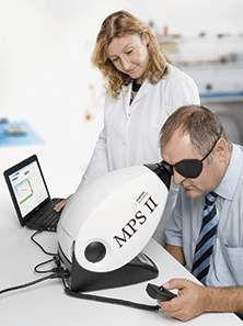
Below, I describe my experience with MP screening, but first, a word about how the technology works. The instrument emits two wavelengths of light that interact with the autofluorescent macular pigment, lipofuscin. One is well absorbed (typically blue, 460 nm), the other is minimally absorbed (typically green, 540 nm). The patient views a small circular stimulus that alternates between the wavelengths. When the frequency of alternation is chosen correctly, the test field appears to flicker. Then the energy of the blue light is adjusted so that the flickering stops. The amount of blue light required to produce this ‘flicker null’ provides a measure of the optical density of MP at that location on the retina and it is recorded when the patient first identifies the flicker. I use an MPS II pigment scanner (Figure 1). Usually, a single, one-minute scan can determine the patient’s MP levels. The scanner delivers a traffic light-style red-amber-green assessment of scan quality – an algorithm assesses blue/green color ratio changes during the scan, and compares this against a normative database of 1.5 million samples. Approximately 90 percent of the scans get a green light; the rest, I repeat.
Useful Tips
In this test, the patient must make reliable judgments about the disappearance of flicker and, as with any visual field examination, practitioner-patient interaction helps prevent noisy data. I recommend the following steps:Before the test
- Take a short patient history, including whether they have had any intraocular lens implantation
- Place a full or reduced aperture trial lens that matches the patient’s reading prescription in the machine’s front slot. If these aren’t available, the instrument has a 4.00 D lens within the optics and helpfully, the HFP procedure is relatively insensitive to blur. Avoid using tinted lenses.
Routine testing
- Occlude the other eye using an eye patch and perform using mesopic or scotopic lighting.
- Measure the right eye, then the left eye – central measurement only – and save the results.
- Tell the patient that they can blink normally, but that the test requires them to concentrate on the task at hand. Make sure the patient understands that when they see flicker on the cetral spot, they need to press the shutter button as quickly as possible. Before performing the main test, do a short (~30 second) familiarization test beforehand to check the speed of their response. The familiarization test also sets the initial blue/green ratio.
- If the patient’s responses are particularly inconsistent during the main test, error messages appear stating “range too high” and “start again”. You will need to restart the scanning procedure.
- When the light scans over the middle spot, the patient will (temporarily) see only black. Some patients think that this is the end of the test. It isn’t. Do not let the patient move their head, as the second test commences when the screen lights up again.
- During the second test, the flickering central light may appear to the patient to be slightly bleached, leaving an after-image. Reassure the patient that this is just because he or she has been staring at it, much like “staring at a bright light for too long”.
- If you see the patient pressing the button too quickly, or losing concentration between responses, pause the test. Repeat the test instructions and decide whether to restart the test immediately, or repeat it later.
- Constantly communicate with the patient using phrases like “you’re doing well”, “look for the flicker’ and “you’re nearly finished”. Silence tends to makes patients question if they are performing the test correctly.
Case Study
Patient GW first presented in March 2010 for a routine eye examination. Drusen were present in both eyes, and fundus photographs were taken. Central vision was present in only one eye due to dense amblyopia, present since birth. She was a non-smoker with no family history of AMD. Her best corrected visual acuity (BCVA) was 6/9 and she experienced glare, particularly when driving at night – even when wearing varifocals with a reflection free coating. Her macular pigmentation optical density (MPOD) was measured at 0.46 (the macular pigment log unit score). She was started on a supplement containing lutein, xeazanthin, omega-3 fatty acids, resveratrol and vitamins and given dietary and exercise advice. On return for a follow-up examination in April 2011, she had fewer drusen, an improved BCVA of 6/6, and glare had resolved. Her macular pigmentation had increased to 0.64, an improvement of over 100 percent. She returned to the clinic in November 2011, where optical coherence tomography (OCT) retinal imaging was performed and revealed drusen on the retinal pigmented epithelium, but no retinal thinning. In November 2012, her MPOD was found to be 0.72 and her visual acuity remained at 6/6 with a similar drusen count and continued resolution of glare.For coexisting pathologies
- Taking central and peripheral measurements is required, if a coexisting pathology, like diabetic maculopathy or AMD, is present. Perform the central measurement first. Inform the patient that they have to stare at the top of the peripheral red target (left target for right eye and vice-versa) and use their side vision to view the central blue flickering target.
- They will want to glance at central blue flickering target, but they must be told firmly to resist this temptation. Staring at the flickering target directly forfeits the result, as it is performing a central measurement again.
- Half-way through the test, pause and get the patient to look at the bottom of the red target from then on, but again only pressing the button when they see the flickering target.
Scott Mackie is an Independent Prescribing Optometrist, Professional Services Director for Visioncall Opticians, Industry Consultant and Visiting Professor at Plymouth University.
