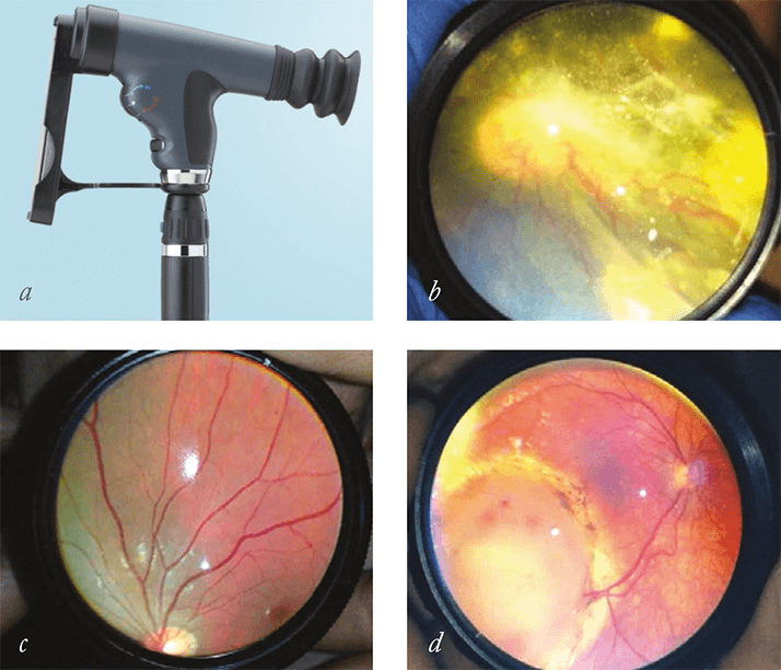
If you think that taking images of the retina requires an ophthalmology clinic decked out in expensive instrumentation – a condensing lens and coaxial light source, a dedicated image acquisition device, all linked to a computer – think again. The rise of smartphones that can shoot high-definition video, take even higher-resolution still images and provide a constant light source from a LED flash, means that everything but the specialized optics can be accomplished with a high-end iPhone or equivalent.
These recent capabilities haven’t escaped the attention of medical diagnostic equipment manufacturers. One, Welch Allyn, lets you pair their €540 PanOptic Ophthalmoscope with an iPhone 4 or 4S with a professional-looking €65 adaptor and its associated free app (Figure 1a). Image acquisition, storage, and distribution are all performed on the smartphone – increasing convenience and cutting costs (the total purchase price excluding the iPhone is only €605). Now, three Boston-based ophthalmologists have lowered the bar even further. Luis Haddock, David Kim and Shizo Mukai of the Massachusetts Eye and Ear Infirmary have come up with a solution that is cheaper and more portable (1). It involves holding a €20, 20 D lens in front of the eye, and using the iPhone to video the images of the lens (Figure 1b–d).
When this has been tried before, results were unsatisfactory, primarily because the in-built Apple iPhone Camera app was poor. Apple’s app did not to allow users to adjust focus or contrast whilst filming, leading to blurry images that were full of glare. Luis, David and Kim’s solution is simple: buy a better camera app. They chose FiLMiC Pro, which currently retails for €2.99 on the Apple iPhone App Store. It enables focus and exposure adjustments during filming. The results are great – both with and without Koeppe lens use, and the group have been able to capture excellent, high-quality fundus images in children under anesthesia and in awake adults. “Our technique provides a simpler and higher-quality method to more consistently produce excellent images of a patient’s fundus,” said Shizuo Mukai, explaining that “this technique has been extremely helpful for us in the Emergency Department setting, in in-patient consultations, and during examinations under anesthesia, as it provides a cheaper and portable option for high-quality fundus-image acquisition for documentation and consultation”. Looking to justify the purchase of the latest smartphone? Upgrading your iPhone is likely to improve the camera in the device – and with it, the quality of your ophthalmoscopy
References
- LJ Haddock, DY Kim, S Mukai, “Simple, Inexpensive Technique for High-Quality Smartphone Fundus Photography in Human and Animal Eyes”, J. Ophthalmol, Article ID 518479 (2013)
