- Worldwide, ROP continues to be a leading cause of childhood blindness, and the traditional standard of care is indirect ophthalmoscopy performed at the bedside
- There are multiple challenges with the current care paradigm for ROP, including time-intensity of the exam, logistical challenges, and high medicolegal liability
- As such, the numbers of skilled ophthalmologists willing to perform ROP examinations are declining, and access to ROP care can be limited
- Telemedicine may improve standards of ROP care, but there are still barriers to overcome
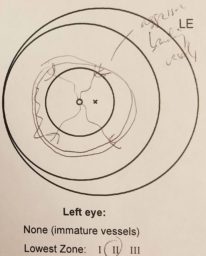
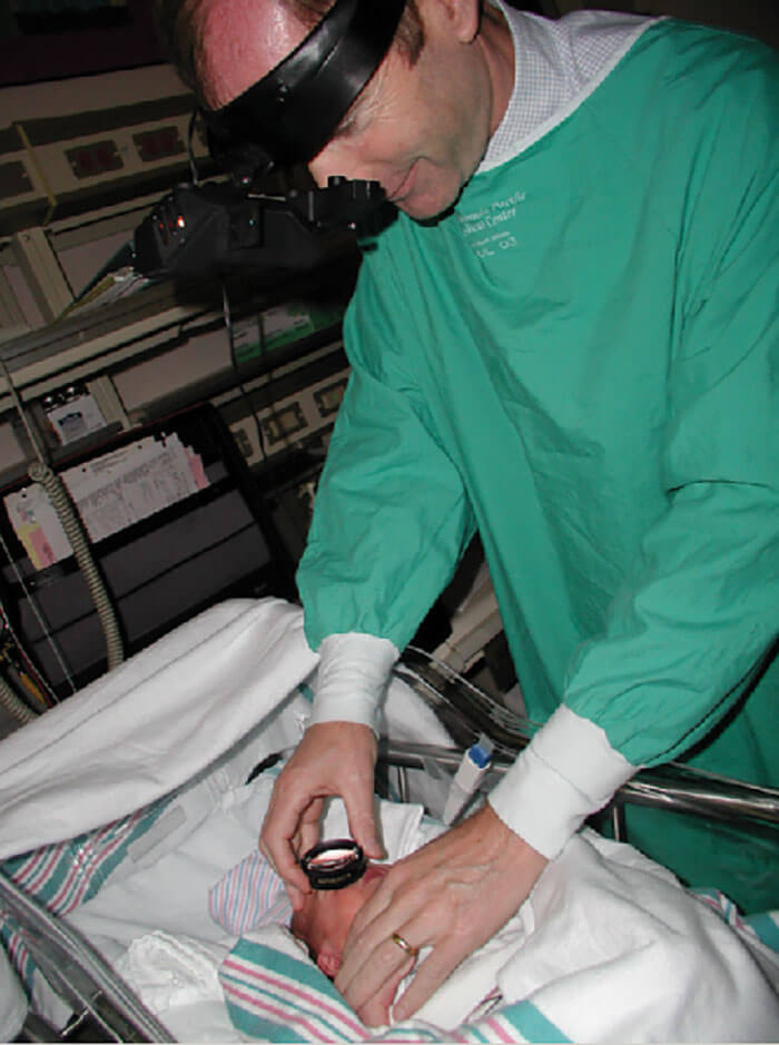
When I started practicing in 2001, the standard of care for the diagnosis of retinopathy of prematurity (ROP) was to perform indirect ophthalmoscopy on premature babies in the neonatal intensive care unit (NICU), and to use those hand-drawn sketches which are familiar to virtually every ophthalmologist (Figure 1, Figure 2). But there are a number of challenges with this care paradigm. Once logistical perspectives have been taken into consideration (traveling time, time taken for coordination between ophthalmologists and neonatologists, and so on), the fact remains that performing ophthalmoscopy on wriggling babies in the NICU is difficult, and hand drawing is inherently an imprecise art that contains a large element of subjectivity. The advances in neonatal care for premature babies over the last six decades have been incredible – babies survive today that are smaller and sicker than were ever thought viable in the past, but these advances have also increased the number of babies at risk of developing ROP. Unfortunately, the number of ophthalmologists who are both trained and willing to assess babies for ROP is decreasing. Why? The reasons include time intensity and the difficulty of the exam, the lack of available adequate training, the complexities of scheduling ROP care, and in the US at least, poor reimbursement. But the elephant in the room is medicolegal liability and the associated high cost of medical indemnity insurance – as far back as 2006, there were reports that many US-based ophthalmologists were considering stopping performing ROP examinations for precisely this reason (1). An unsurprising consequence is the reduced availability of ROP care, particularly in rural and medically under-served areas. Could telemedicine provide the much-needed solution?
A telemedicine solution?
In addition to the challenges mentioned above, the accurate diagnosis of ROP can be hindered by the significant variability among different ophthalmologists. In the US, half of all clinicians performing ROP exams are general ophthalmologists without retinal or pediatric specialty training (2) – but even so, clinically significant ROP is still being missed in some cases by retinal specialists and pediatric ophthalmologists (Figure 3) (3, 4), and it is clear that there are a number of gaps in the training for and the delivery of quality ROP care. Using the telemedicine approach, NICU staff are trained to image the retina of at-risk infants using wide-angle digital retinal photography. Those pictures are then be sent for remote diagnosis by experts, and babies who then need a full eye exam – based on their retinal pictures – can be referred to experts at a central location. The potential benefits for ROP care with this approach are many. Improved quality, accessibility and cost-effectiveness of ROP care; the captured photographs should allow ROP diagnosis to be more objective relative to hand-drawn sketches, and the approach should also improve the monitoring of the disease.
In the real world
There are many published ROP telemedicine studies, and based on a technology review of 10 of these, telemedicine demonstrates both a high sensitivity and a high specificity for diagnosing clinically significant – that is treatment-requiring or referral-warranted – ROP (5). More recently, a large-scale trial involving over 1,200 babies has shown a high sensitivity for diagnosing clinically significant ROP (NCT01264276). In this study, premature infants from 13 North American centers were assessed by both diagnostic examinations from ophthalmologists, and remotely by non-physician graders using digital imaging. The sensitivity of remote grading in identifying referral-warranted ROP was 82–90 percent when one eye was examined and 90 percent when both eyes were examined (6). So it seems that a photograph can feasibly allow a correct diagnosis to be drawn. But all of these studies were based on the assumption that indirect ophthalmoscopy is the gold standard for ROP diagnosis. This leads us to question “What is the correct diagnosis for ROP? And how do we know that ophthalmoscopy is inherently better than looking at a photo of the eye?” This is a difficult question to answer, but we started by looking at intraphysician agreement by pediatric ophthalmologists who regularly performed ophthalmoscopic exams for ROP diagnosis. Six months after the initial ophthalmoscopic examination we asked them to examine retinal photos which had been taken at the time of the examination, and we found an 86 percent intraphysician agreement between the ophthalmoscopic diagnosis and the telemedicine diagnosis. Despite a high-level of agreement between the two diagnoses, it still meant that in 14 percent of cases there were discrepancies in diagnosis by the same person. What went wrong? It seems that in 32 percent of the cases, the photos showed something that appeared to have been missed in the original ophthalmoscopic exam (in which no disease had been identified). In 29 percent of these cases, there were discrepancies about subtype of disease: zone I disease was identified by telemedicine and zone II by ophthalmoscopy, or vice versa. In principle, telemedicine might allow for a more accurate diagnosis: images can be closely scrutinized more effectively as opposed to doing this discrimination on a wiggling baby at the bedside. Telemedicine is already being used in the real-world. Currently in the US, there are large-scale real-world telemedicine programs – some of which have been operational for almost a decade (7, 8). India has a well-established telemedicine program – KIDROP – which has been operational for over eight years. Promisingly, the 2013 revision to the American Academy of Ophthalmology and American Academy of Pediatrics (AAO-AAP) joint ROP screening guidelines acknowledge the use of telemedicine for ROP diagnosis (9), and both societies have jointly provided guidance on the use of telemedicine for the diagnosis of ROP (10). The use of telemedicine in a real-world setting might also support the use of computer-based image analysis tools, such as ROPtool and FocusROP, to accurately measure disease and monitor progression, and we and others – such as David Wallace – have done work in this area.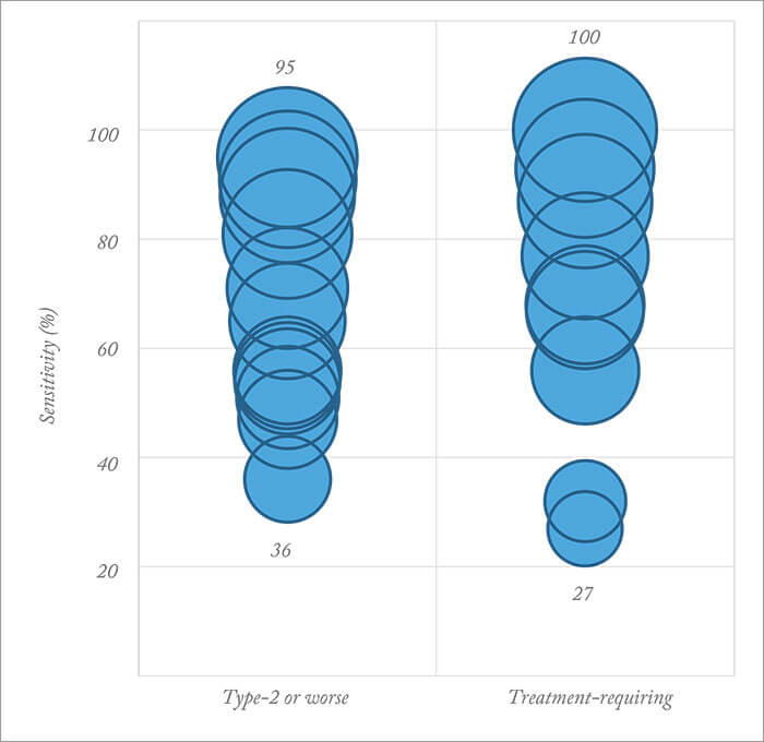
Barriers remain
The fact that there are telemedicine programs operating now suggests that it is feasible in the real-world, but a few barriers remain. We need to develop effective training programs for NICU nurses and staff in order to teach them how to take the pictures and how to interpret them. There is the issue of workflow – we need to get these systems to work in the real-world. Reading software, assignment of individual roles and responsibilities for ophthalmologists and neonatologists, criteria for identification of inadequate images, and transfer rules all need to be defined. In addition, policies for licensure, insurance cover and reimbursement all need to be considered – particularly in the US. Most importantly, we need to challenge the perception that indirect ophthalmoscopy is the gold standard for ROP diagnosis. Telemedicine will bring ROP care into the digital area and improve the standard of ROP care, but we need to develop large scale solutions to overcome the remaining barriers for telemedicine. In some ways, I believe that overcoming these remaining barriers is more of a political or economic issue rather than a medical or technological one.References
- K. Altersitz, “Survey: Physicians being driven away from ROP treatment”, Ocular Surgery News U.S. Edition (2006). Accessed April 11, 2016. AR Kemper et al., “Retinopathy of prematurity care: patterns of care and workforce analysis”, J AAPOS, 12, 344–348 (2008). PMID: 18440256. RVP Chan et al., “Accuracy of retinopathy of prematurity diagnosis by retinal fellows”, Retina, 30, 958–965 (2010). PMID: 20168274. JS Myung et al., “Accuracy of retinopathy of prematurity image-based diagnosis by pediatric ophthalmology fellows: implications for training”, J AAPOS, 15, 573–578 (2011). PMID: 22153403. MF Chiang et al., “Detection of clinically significant retinopathy of prematurity using wide-angle digital retinal photography: a report by the American Academy of Ophthalmology”, Ophthalmol, 119, 1272–1280 (2012). PMID: 22541632. GE Quinn et al., “Validity of a telemedicine system for the evaluation of acute-phase retinopathy of prematurity”, JAMA Ophthalmol, 132, 1178–1184 (2014). PMID: 24970095. SK Wang et al., “SUNDROP: six years of screening retinopathy of prematurity with telemedicine”, Can J Ophthalmol, 50, 101–106 (2015). PMID: 25863848. DT Weaver and TJ Murdock,“Telemedicinedetection of type 1 ROP in a distant neonatal intensive care unit”, J AAPOS, 16, 229–233 (2012). PMID: 22681938. WM Fierson et al., “Screening examination of premature infants for retinopathy of prematurity”, Pediatr, 131, 189–195(2013). PMID: 23277315. WM Fierson et al “Telemedicine for evaluation for retinopathy of prematurity”, Pediatr, 135, e238–254(2015). PMID: 25548330.
