Conexiant
Login
  • Corneal Physician
  • Glaucoma Physician
  • New Retinal Physician
  • Ophthalmology Management
  • Ophthalmic Professional
  • Presbyopia Physician
  • Retinal Physician
The Ophthalmologist
  • Explore

    Explore

    • Latest
    • Insights
    • Case Studies
    • Opinion & Personal Narratives
    • Research & Innovations
    • Product Profiles

    Featured Topics

    • Anterior Segment
    • Glaucoma
    • Retina

    Issues

    • Latest Issue
    • Archive
  • Subspecialties
    • Cataract
    • Cornea
    • Glaucoma
    • Neuro-ophthalmology
    • Oculoplastics
    • Pediatric
    • Retina
  • Business

    Business & Profession

    • Professional Development
    • Business and Entrepreneurship
    • Practice Management
    • Health Economics & Policy
  • Training & Education

    Career Development

    • Professional Development
    • Career Pathways

    Events

    • Webinars
    • Live Events
  • Events
    • Live Events
    • Webinars
  • Community

    People & Profiles

    • Power List
    • Voices in the Community
    • Authors & Contributors
  • Multimedia
    • Video
    • Podcasts
Subscribe
Subscribe

False

Advertisement
The Ophthalmologist / Issues / 2014 / Jan / Retinal Visualization of Schizophrenia

Retinal Visualization of Schizophrenia

OCT imaging visualizes retinal nerve fiber layers and the thickness of the macula; abnormalities in either can predict the stage and duration of schizophrenia.

By Mark Hillen 1/20/2014 1 min read

Share

0114-204-maim-img

Patients with schizophrenia exhibit a progressive reduction in brain volume as the disease advances. From the very first schizophrenic episode, during the prodromal phase and even in those with a high genetic risk of developing the disease, volume deficits in grey matter are detectable.

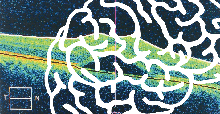
When researchers at the Department of Ophthalmology at the University of Malaya, in Kuala Lumpur, Malaysia, decided to see if these abnormalities could be visualized in the retina by spectral-domain optical coherence tomography (SD-OCT) they found that, relative to age-matched controls, patients with schizophrenia exhibited significant thinning of the retinal nerve fiber layer (RNFL) and the macula; overall macular volume was reduced too. The study revealed significant inverse correlations between the duration of schizophrenic illness and peripapillary RNFL thickness (r=–0.36), macula thickness (r=–0.38), and macula volume reduction (r=–0.36).

The authors concluded that “SD-OCT can be a useful tool for diagnosis and monitoring the progression of this disease.” As we’ve previously reported, the retinal vasculature can also predict the status of systemic diseases like diabetes [0113-301] – even without injecting β-cells from the Islets of Langerhans into the anterior chamber (this issue). It seems that either ophthalmologists will increasingly assess non-ophthalmic disease states in patients or that the practitioners of other specialties will soon be investing in imaging equipment that was once solely the domain of eye care specialists.

About the Author(s)

Mark Hillen

I spent seven years as a medical writer, writing primary and review manuscripts, congress presentations and marketing materials for numerous – and mostly German – pharmaceutical companies. Prior to my adventures in medical communications, I was a Wellcome Trust PhD student at the University of Edinburgh.

More Articles by Mark Hillen

Related Content

Newsletters

Receive the latest Ophthalmology news, personalities, education, and career development – weekly to your inbox.

Newsletter Signup Image

False

Advertisement

False

Advertisement

Explore More in Ophthalmology

Dive deeper into the world of Ophthalmology. Explore the latest articles, case studies, expert insights, and groundbreaking research.

False

Advertisement
The Ophthalmologist
Subscribe

About

  • About Us
  • Work at Conexiant Europe
  • Terms and Conditions
  • Privacy Policy
  • Advertise With Us
  • Contact Us

Copyright © 2025 Texere Publishing Limited (trading as Conexiant), with registered number 08113419 whose registered office is at Booths No. 1, Booths Park, Chelford Road, Knutsford, England, WA16 8GS.

Disclaimer

The Ophthalmologist website is intended solely for the eyes of healthcare professionals. Please confirm below: