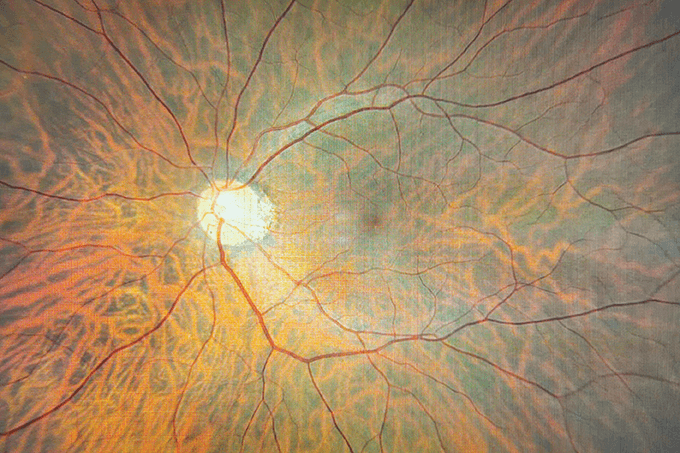
Baby blue. A multinational study led by researchers at the University of Leicester, UK, has deciphered how genetics influence visual development in developing babies’ eyes – more specifically looking at genes associated with arrested development of the fovea (1). They investigated a cohort of almost one thousand people with confirmed genetic disorders associated with foveal hypoplasia - classifying how foveal development correlates to genotype and the visual outcome.
Extending family. NEI researchers have identified a novel early-onset macular dystrophy that occurs as a result of new mutations in the gene TIMP3, already known to be associated with the disease (2). These new mutations were not in the mature protein, but in the signal peptide preventing the immature protein from being cleaved.
Marked targets. Fibroblasts from primary open angle glaucoma patients were reprogrammed into stem cells and differentiated to form a model of the retina and optic nerve to identify unknown genetic markers of glaucoma (3). 312 genetic variants were associated with the target retinal cells and 97 genetic clusters were linked to glaucoma caused damage.
Pixel by pixel. A machine learning model uses a pixel stratification approach to comprehensively reconstruct and examine the vitreous anatomy in 3D. The technique produces high quality 3D movies, similar to triamcinolone-assisted vitrectomy or postmortem dye injection (4). By examining the vitreous structure beyond the vitreoretinal interface there are applications for many macular diseases and diabetic retinopathy. The approach uses swept source OCT (SS-OCT) scans that are analyzed through Fiji (is just image J) to stack into cubic voxels. After processing the signals from the vitreous gel, spaces within, and interfaces between them, two classes of “Septa” and “Other” were defined - these pixels were assigned to either of the classes, which in turn trained the classifier system.
Intelligent reclassification. A new proposed framework for classifying diabetic retinopathy uses OCT and OCT-A with AI to provide specialized classification, all under one imaging modality (5).
Safe but inefficient. An early LHON clinical trial shows that gene therapy treatment trial is safe but not effective at improving or slowing vision loss (6).
Plateau in progress. Although visual impairment in adults with diabetes was in decline since 2000, this may have plateaued from 2012 (7). The study performed by the CDC provides insights into vision impairment in adults with diabetes.
Through thick and thin. Retinal nerve fiber layer thickness is associated with cognitive function and subsequent cognitive decline (8). This information could be applied to OCT eye tests as a predictive biomarker for cognitive function in older patients.
References
- HJ Kuht et al., Ophthalmology, 129, 708 (2022). PMID: 35157951.
- B Guan et al., JAMA Ophthalmol, 140, 730 (2022). PMID: 35679059.
- M Daniszewski et al., Cell Genomics, 2, 8 (2022). Doi: 10.1016/j.xgen.2022.100142.
- A Thi et al., Transl Vis Sci Technol, 11, 3 (2022). PMID: 35802368.
- P Zang et al., Transl Vis Sci Technol, 11, 10 (2022). PMID: 35822949.
- BL Lam et al., Am J Ophthalmol, S0002-9394, 00088 (2022). PMID: 35271811.
- EA Lundeen et al., Poster Presentation at ADA (2022). Available at: https://bit.ly/3z9dRuv.
- HM Kim et al., JAMA Ophthalmol, 140, 683 (2022). PMID: 35616950.
