Conexiant
Login
  • Corneal Physician
  • Glaucoma Physician
  • New Retinal Physician
  • Ophthalmology Management
  • Ophthalmic Professional
  • Presbyopia Physician
  • Retinal Physician
The Ophthalmologist
  • Explore

    Explore

    • Latest
    • Insights
    • Case Studies
    • Opinion & Personal Narratives
    • Research & Innovations
    • Product Profiles

    Featured Topics

    • Anterior Segment
    • Glaucoma
    • Retina

    Issues

    • Latest Issue
    • Archive
  • Subspecialties
    • Cataract
    • Cornea
    • Glaucoma
    • Neuro-ophthalmology
    • Oculoplastics
    • Pediatric
    • Retina
  • Business

    Business & Profession

    • Professional Development
    • Business and Entrepreneurship
    • Practice Management
    • Health Economics & Policy
  • Training & Education

    Career Development

    • Professional Development
    • Career Pathways

    Events

    • Webinars
    • Live Events
  • Events
    • Live Events
    • Webinars
  • Community

    People & Profiles

    • Power List
    • Voices in the Community
    • Authors & Contributors
  • Multimedia
    • Video
    • Podcasts
Subscribe
Subscribe

False

Advertisement
The Ophthalmologist / Issues / 2018 / Jun / Research
Retina Research & Innovations

Research

By Ruth Steer 6/27/2018 1 min read

Share

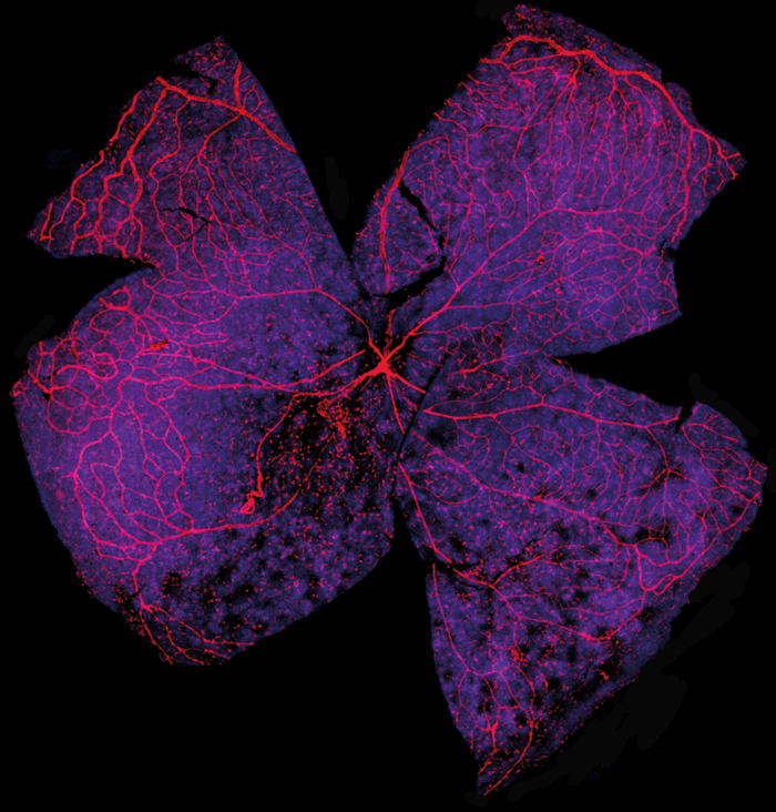
A normal mouse retinal flatmount depicting the optic nerve showing the retinal vasculature stained with isolectin (red) and GFAP (green) and DAPI.

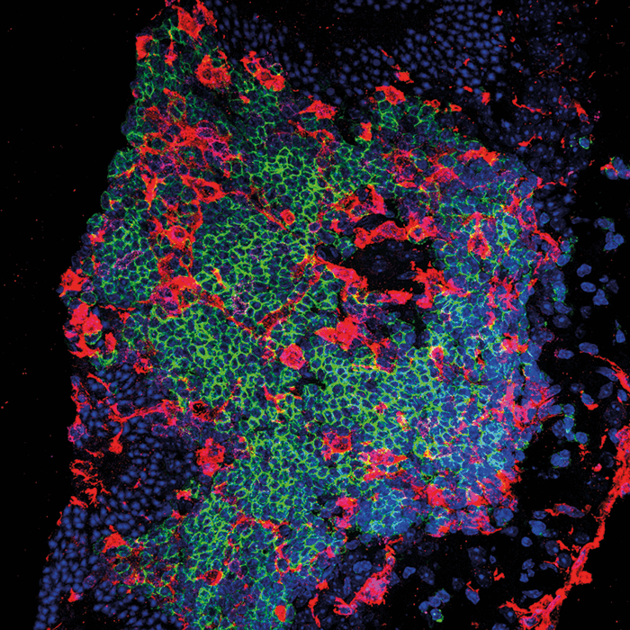
A tertiary retinal lymphoid aggregate in a mouse with spontaneous uveitis. The staining shows the presence on microglia (red), B cells (green), and T cells (magenta) and DAPI.

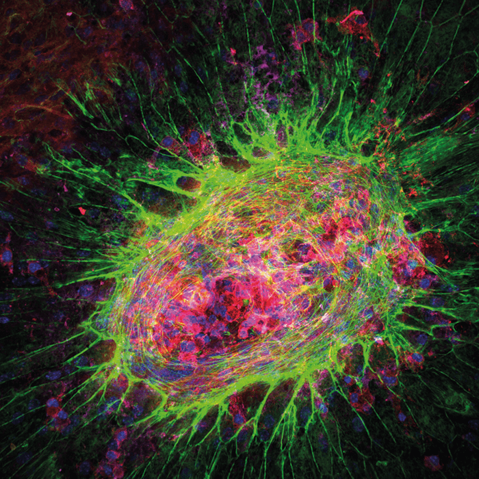
A mouse RPE flatmount with laser injury showing choroidal neovascularization. Staining shows phalloidin (RPE stained), isolectin (blood vessels), and DAPI.

A  mouse retinal flatmount showing the retinal vasculature stained for isolectin (red) and DAPI. The mouse was virally infected with Zika virus.

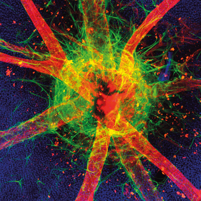
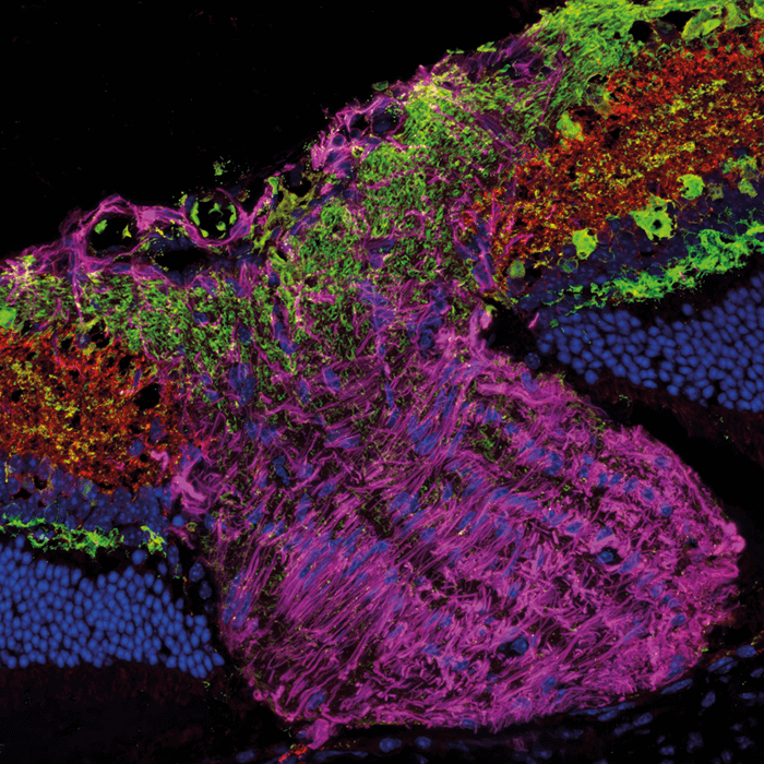
A normal mouse optic nerve head stained for synapsin, PKC-A, and GFAP and DAPI. This selection of images were contributed by Jennifer Kielczewski, a Staff Scientist with the National Eye Institute, Biological Imaging Core (BIC) in Bethesda, MD, USA. Her research interests include using imaging techniques to study retinal pathologies, ocular inflammation, and neuronal degeneration in diseased animal models.

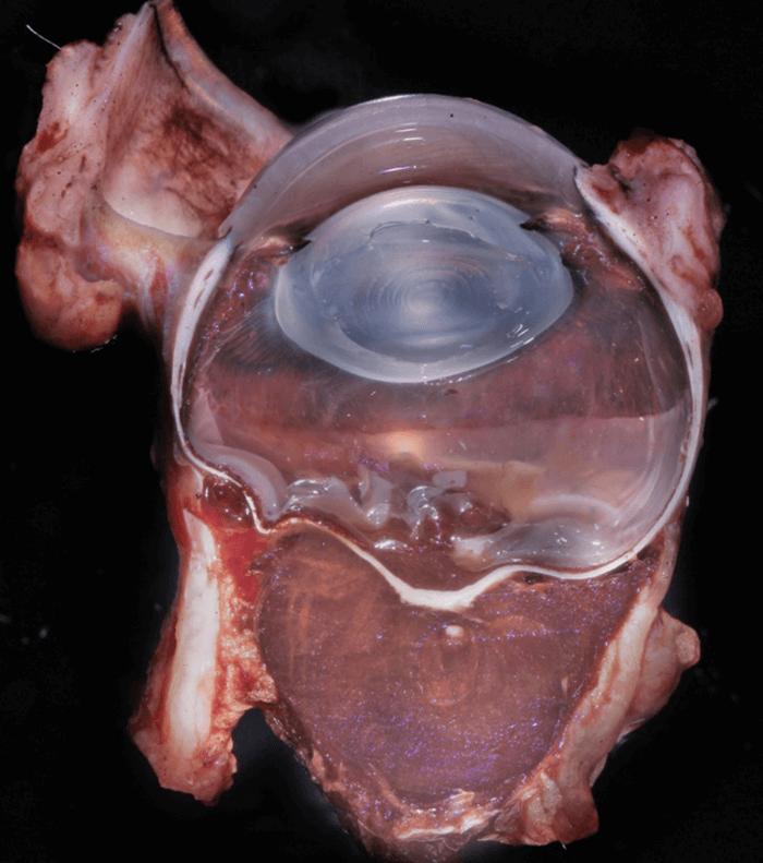
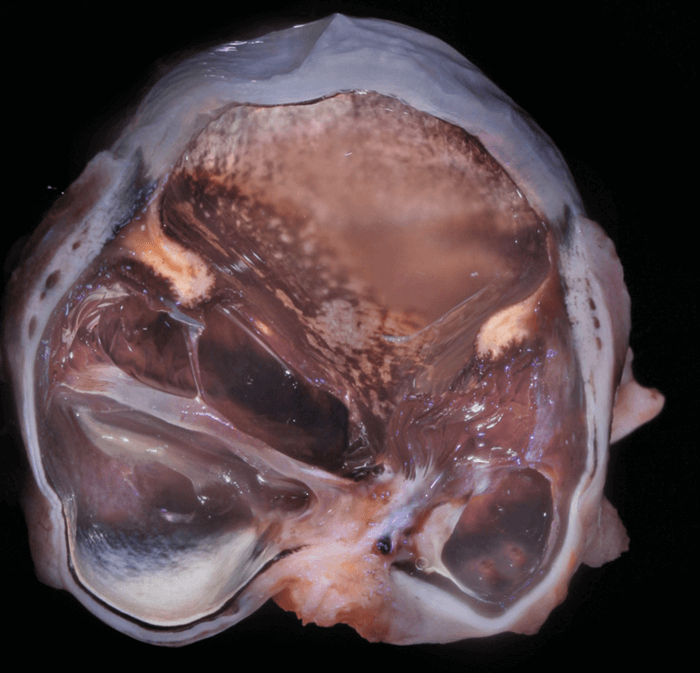
This is the globe of an adult cat with posterior scleral rupture due to presumed blunt trauma. The anterior chamber is collapsed and the pigmented iridal tissue lines the posterior cornea. Pigmented uveal tissue (likely ciliary body) stretches backwards and contacts fibrotic tissue at the posterior scleral rupture site. The lens capsule was ruptured and only recognized microscopically associated with the fibrotic tissue at the site of the posterior scleral rupture.

In this canine globe, a choroidal melanocytoma extended through the posterior sclera and formed a mass behind the globe. Though locally destructive, this type of benign melanocytic tumor rarely metastasizes. The Comparative Ocular Pathology Laboratory of Wisconsin (COPLOW), USA. Further images can be found on the COPLOW Facebook page: bit.ly/COPLOW.

Art of Ophthalmology
Research
Global Ophthalmology
In The clinic

About the Author(s)

Ruth Steer

More Articles by Ruth Steer

Related Content

Newsletters

Receive the latest Ophthalmology news, personalities, education, and career development – weekly to your inbox.

Newsletter Signup Image

False

Advertisement

False

Advertisement

Explore More in Ophthalmology

Dive deeper into the world of Ophthalmology. Explore the latest articles, case studies, expert insights, and groundbreaking research.

False

Advertisement
The Ophthalmologist
Subscribe

About

  • About Us
  • Work at Conexiant Europe
  • Terms and Conditions
  • Privacy Policy
  • Advertise With Us
  • Contact Us

Copyright © 2025 Texere Publishing Limited (trading as Conexiant), with registered number 08113419 whose registered office is at Booths No. 1, Booths Park, Chelford Road, Knutsford, England, WA16 8GS.

Disclaimer

The Ophthalmologist website is intended solely for the eyes of healthcare professionals. Please confirm below: