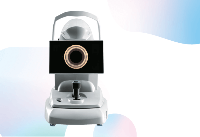
The solution to limitations in iridocorneal angle imaging is finally here. Introducing the NIDEK GS-1 – the first automated gonioscopy device that can instantly document the iridocorneal angle in real-color. Simple to use and more definitive than subjective pathology sketches, GS-1’s high-resolution goniophotographs not only enhance the quality of assessment, but allow for more comprehensive follow-up.
The GS-1 automatically acquires color photographs of the iridocorneal angle using an exclusive 16-mirror goniolens, specifically designed by NIDEK. The internal optical system automatically rotates and acquires color photographs of the iridocorneal angle in 16 directions, documenting the entire angle. Each direction can be captured in 17 different foci, which brings true versatility to iridocorneal angle photography.
The resulting 360-degree images can be displayed as a single shot with circular or linear stitching, allowing localization of features and pathologies. The resulting high-resolution color images can then be exported in JPEG, PNG and PDF files so they can be easily shared with the patient or fellow clinicians. Built with patient comfort in mind, the gel-assisted soft-contact interface ensures that the multimirror prism does not touch the cornea.
Given gonioscopy’s crucial role in retinal disease and glaucoma – both of which require a thorough understanding of the iridocorneal angle anatomy, it is surprising that clinicians typically leave handwritten notes or sketches in the patient chart. Though some digital imaging devices can be used – anterior OCT and ultrasound biomicroscopy, for example – neither provide a picture as clear or definitive as a detailed color image of iridocorneal anatomy.
More than that, the skills needed to properly perform gonioscopic examination have limited its use in the daily practice. The NIDEK GS-1 is born of need and therefore provides a perfect solution by simplifying the process so that it can be performed by doctors and technicians, increasing screening efficacy. Thus, the device frees up time for the clinician to assess and plan treatment, knowing that they have the convenience of re-assessing the angle via the digital goniophotographs at any time.
However, the practical applications of the GS-1 go beyond just the advanced analysis of iridocorneal angle, the device can also be used for early glaucoma screening, patient education, and as a means of supporting MIGS device analysis, assessment and follow-up. Though technological innovation has revolutionized almost all aspects of ophthalmology, gonioscopy has remained virtually unchanged for decades. The NIDEK GS-1 was born of need.
