Conexiant
Login
  • Corneal Physician
  • Glaucoma Physician
  • New Retinal Physician
  • Ophthalmology Management
  • Ophthalmic Professional
  • Presbyopia Physician
  • Retinal Physician
The Ophthalmologist
  • Explore

    Explore

    • Latest
    • Insights
    • Case Studies
    • Opinion & Personal Narratives
    • Research & Innovations
    • Product Profiles

    Featured Topics

    • Anterior Segment
    • Glaucoma
    • Retina

    Issues

    • Latest Issue
    • Archive
  • Subspecialties
    • Cataract
    • Cornea
    • Glaucoma
    • Neuro-ophthalmology
    • Oculoplastics
    • Optometry
    • Pediatric
    • Retina
  • Business

    Business & Profession

    • Professional Development
    • Business and Entrepreneurship
    • Practice Management
    • Health Economics & Policy
  • Training & Education

    Career Development

    • Professional Development
    • Career Pathways

    Events

    • Webinars
    • Live Events
  • Events
    • Live Events
    • Webinars
  • Community

    People & Profiles

    • Power List
    • Voices in the Community
    • Authors & Contributors
  • Multimedia
    • Video
    • Podcasts
Subscribe
Subscribe

False

Advertisement
The Ophthalmologist / Issues / 2023 / Mar / Printing for Progress
Research & Innovations Retina

Printing for Progress

How 3D bioprinted outer blood-retina barrier could boost AMD research – and even provide transplantable tissue

By Sarah Healey 3/13/2023 2 min read

Share

Credit: Collage images sourced from Shutterstock and Unsplash.com

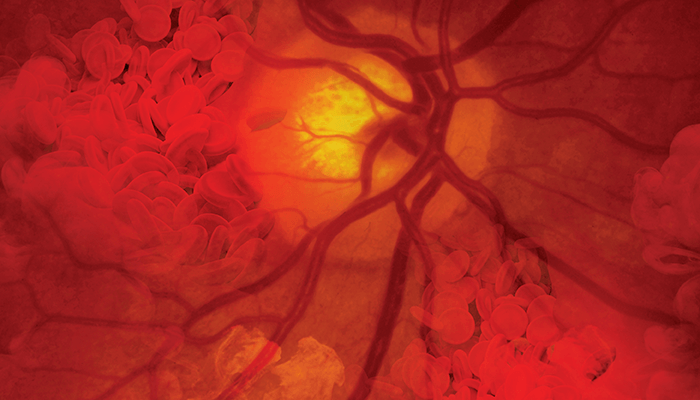
Although it is known that age-related macular degeneration (AMD) develops in the outer blood-retina barrier (oBRB), there remains a lack of understanding around the mechanisms of AMD initiation and progression to advanced dry and wet stages. The short supply of physiologically relevant human oBRB models is one factor that preventing advanced research into degenerative retinal disease, so a team from the National Eye Institute (NEI) set out to engineer their own.

To do this, researchers engineered a native-like three-dimensional (3D) oBRB tissue (3D-oBRB) by bioprinting endothelial cells, pericytes, and fibroblasts on the basal side of a biodegradable scaffold and establishing an RPE monolayer on top. In this 3D-oBRB model, a fully-polarized RPE monolayer provides barrier resistance, induces choriocapillaris fenestration, and supports the formation of Bruch's-membrane-like structure by inducing changes in gene expression in cells of the choroid (1).

Khapil Bharti, the leader of the NEI Section on Ocular and Stem Cell Translational Research, reports that the tissue took four weeks to mature, by which time the RPE cells had formed a confluent polarized monolayer and the scaffold had degraded completely, leaving behind a Bruch’s membrane-like structure. On the other side of Bruch’s membrane, fibroblasts formed a matrix within which endothelial cells and pericytes formed capillaries (similar to native choriocapillaris). This tissue behaved similarly to native RPE-choriocapillaris (CC) tissue and was able to reproduce key cellular phenotypes of AMD, cementing 3D-oBRB as an important model for studying the progression of AMD.

As a result of all the cell types of the 3D-oBRB being induced from patient derived, pluripotent stem cells, researchers are now able to re-create patient disease in a dish, enabling earlier disease detection and intervention research. In addition, Bharti says: ‘this tissue can be used to test the safety and efficacy of new drugs replacing animal usage for drug testing. In the future, we can also use such tissues for transplantation in patients to replace degenerated and damaged retina.’

Bharti and his collaborators are confident that the 3D-oBRB can provide a platform to address previously unanswered questions, such as how exosomes and cytokines regulate RPE–choroid crosstalk. Other areas for further research include the pathological effects of drusen accumulating under the RPE, functionality of the RPE, and choriocapillaris fenestration.

References

  1. M J Song et al., “Bioprinted 3D outer retina barrier uncovers RPE-dependent choroidal phenotype in advanced macular degeneration,” Nat Methods, [Online ahead of print] (2022). PMID: 36550275.

About the Author(s)

Sarah Healey

Communicating stories in a way that is accessible to all was one of the focal points of my Creative Writing degree. Although writing magical realism is a fun endeavor (and one I still dabble in), getting to the heart of human stories has always been the driving motivator behind my writing. At Texere, I am able to connect with the people behind scientific breakthroughs and share their stories in a way that is impactful and engaging.

More Articles by Sarah Healey

Related Content

Newsletters

Receive the latest Ophthalmology news, personalities, education, and career development – weekly to your inbox.

Newsletter Signup Image

False

Advertisement

False

Advertisement

Explore More in Ophthalmology

Dive deeper into the world of Ophthalmology. Explore the latest articles, case studies, expert insights, and groundbreaking research.

False

Advertisement
The Ophthalmologist
Subscribe

About

  • About Us
  • Work at Conexiant Europe
  • Terms and Conditions
  • Privacy Policy
  • Advertise With Us
  • Contact Us

Copyright © 2025 Texere Publishing Limited (trading as Conexiant), with registered number 08113419 whose registered office is at Booths No. 1, Booths Park, Chelford Road, Knutsford, England, WA16 8GS.

Disclaimer

The Ophthalmologist website is intended solely for the eyes of healthcare professionals. Please confirm below: