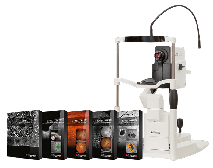
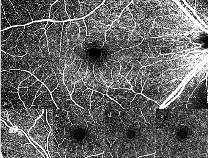
The SPECTRALIS® SD-OCT is Heidelberg Engineering’s expandable diagnostic imaging platform which combines scanning laser fundus imaging with high-resolution OCT. It can be configured according to different diagnostic workflows with options including OCT, multiple scanning laser fundus imaging modalities, widefield and ultra-widefield modules, scanning laser angiography and OCT angiography (OCTA). The OCT Angiography Module* is the latest addition to the SPECTRALIS platform and offers three-dimensional insights into vascular flow, with a flexible combination of non-invasive high-resolution imaging and a high-speed widefield view (see image a). The SPECTRALIS OCT Angiography Module delivers high-resolution OCTA images depicting fine capillary networks in great detail and a precise segmentation of all four histologically-validated retinal vascular plexuses (see images b-e) with minimal motion artifacts. The module offers the flexibility to customize the slab position on every B-scan image, allowing for visualization of pathology in any retinal layer. The dynamic image fusion of OCT and OCTA supports precise localization of flow in abnormal vessels. The SPECTRALIS also enables a unique hybrid approach to angiography with a precise, pixel-to-pixel correlation of OCTA follow-ups on existing FA and ICGA images. In short, the SPECTRALIS OCT Angiography Module provides the dynamic tools needed to master the application and interpretation of this novel imaging modality. It combines OCTA with structural OCT and fundus imaging resulting in a powerful multimodal approach to support clinical assessment and treatment decisions.
The SPECTRALIS OCT Angiography Module provides high-resolution OCTA images with a lateral resolution of 5.7 μm per pixel for the visualization of capillary vessels. The axial resolution of 3.9 μm per pixel enables detailed retinal layer segmentation. All four vascular networks can then be examined: vasculature in the nerve fiber layer (b), in the ganglion cell layer (c), at the border of the inner plexiform layer to the inner nuclear layer (d), and at the border of the inner nuclear layer to the outer plexiform layer (e).
*The OCT Angiography Module is available for purchase only outside the United States.
