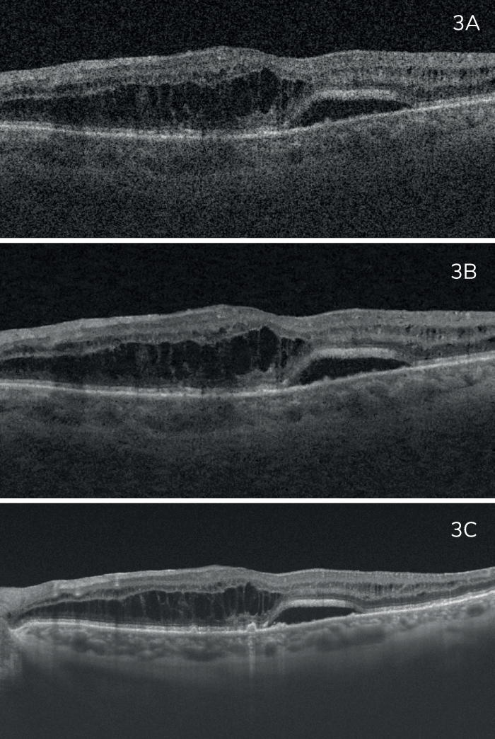
Above: Wet age-related macular degeneration, Triton 3D OCT scan, PixelSmart image. Image courtesy of Heloísa Nascimento.
Topcon Healthcare, an Optical Coherence Tomography (OCT) instrumentation leader, offers both spectral domain (SD) and swept-source (SS) OCT options, has introduced a new image processing algorithm to its unique DRI OCT Triton™ series. The novel PixelSmart technology takes SS-OCT imaging to the next level and offers unparalleled image quality by reducing speckle noise and improving image contrast.
Many OCT devices repeatedly scan each retinal location and average the data, in order to obtain satisfactory image quality. Clinicians using such devices will be excited to try PixelSmart which can provide a detailed assessment of the posterior segment both quickly and efficiently. Thanks to Triton’s exceptional high-density SS-OCT data, the new image processing algorithm is capable of producing rich, detailed images without compromising speed or the scan area.
Here, ophthalmology experts specializing in retinal care present clinical cases assessed with the Triton utilizing PixelSmart.
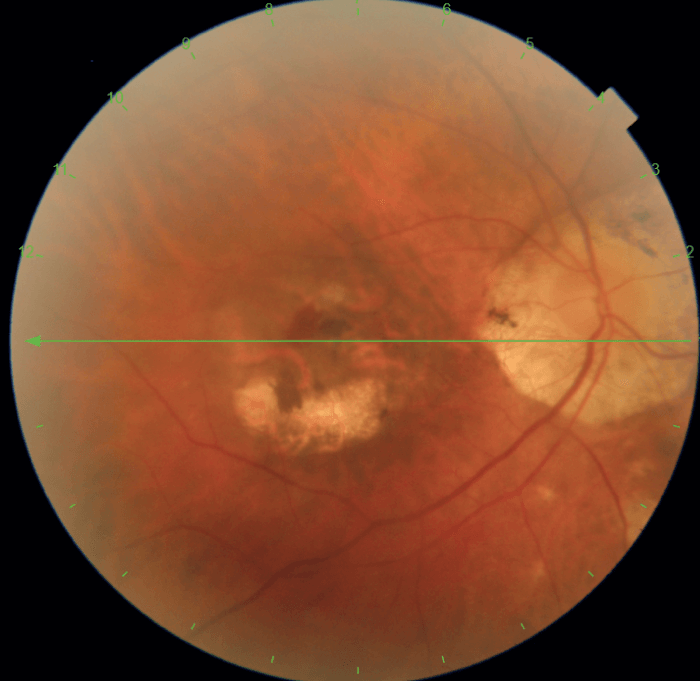
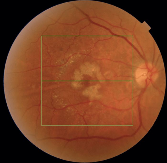
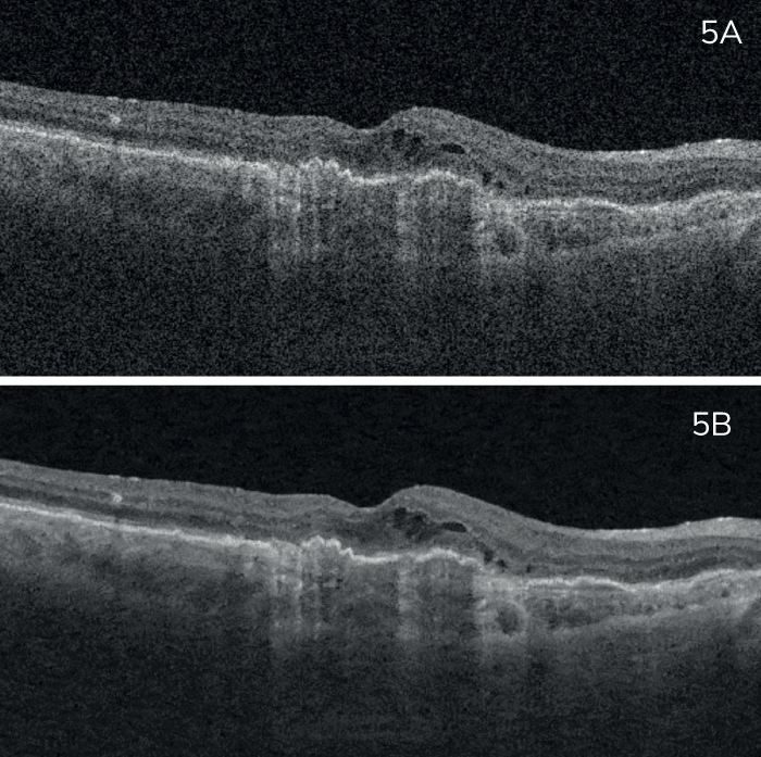
Luis Arias, MD, PhD, Head of the Retina Department of University Hospital of Bellvitge and Aggregate Professor of Ophthalmology, University of Barcelona, Spain, presents a case of a 66-year-old male patient suffering from high myopia and a choroidal neovascular membrane, with a previous history of photodynamic treatment, 20 years prior.
Professor Arias shares his view on the utility of the new image processing technology available from Topcon Healthcare, “With a good compromise between acquisition time and image quality, PixelSmart can improve workflow in busy clinics. Within a couple of seconds, I can analyze high density scans with good image quality, allowing me to evaluate details of the vitreous, retina and choroid.”
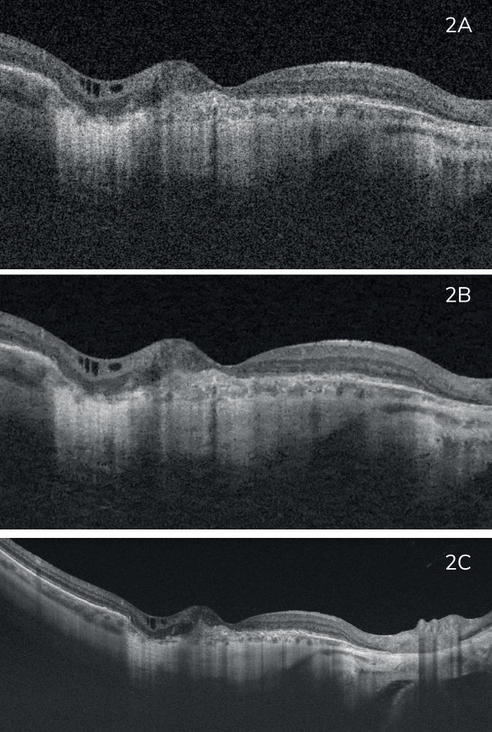
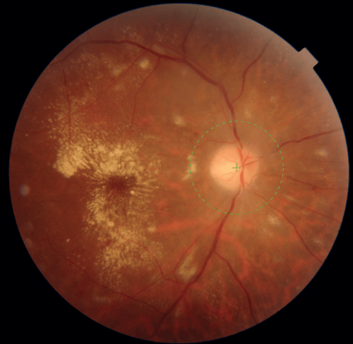
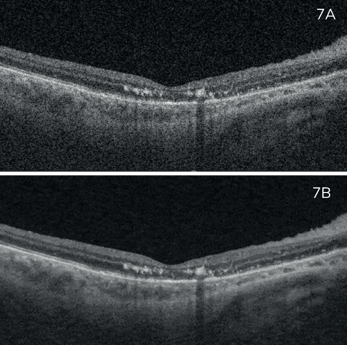
José Maria Ruiz-Moreno, MD, PhD, Head of Ophthalmology, Puerta de Hierro University Hospital, Majadahonda, Madrid, Professor at the University of Castilla-La Mancha Medical School, Albacete, Spain, presents a case of a 77-year-old female patient with diabetic macular edema.
On the novel imaging algorithm, Professor Moreno comments, “PixelSmart is a great tool for screening the retina. It improves the image quality of standard 3D scans, aiding in the assessment of retinal diseases.”

Heloísa Nascimento, MD, PhD, from the Department of Ophthalmology, Federal University of São Paulo, Paulista Medical School – UNIFESP/EPM, Brazil, presents a case study of an 80-year-old female patient with wet age-related macular degeneration.
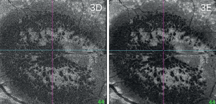
Heloísa Nascimento, MD, PhD, also shares a case study of a 33-year-old male patient diagnosed with ocular syphilis.
Professor Nascimento shares her impressions of Topcon Heathcare’s new image processing technology, “PixelSmart has greatly improved the image quality of 3D scans. Having good images with a high-density scan allows for a better evaluation of the retinal and choroidal alterations. This is especially important to image patients who do not collaborate with the exam or have poor fixation, when it is difficult to position a line or radial high-quality scan exactly where the alteration is.”
Blink-of-an-eye imaging
With PixelSmart technology, Topcon Healthcare once again pushes the boundaries of OCT imaging, providing unprecedented image quality available to clinicians for instant, precise analysis, improving the clinic’s performance and – ultimately – patients’ outcomes. It further cements Topcon Healthcare’s position as the market leader in the field of OCT, constantly innovating and improving its portfolio to deliver the best solutions to ophthalmic practices around the world.
Topcon Healthcare recommends installing the viewing system with PixelSmart on a network to allow image review from exams rooms in addition to the imaging room.
Not all products, services or offers are approved or offered in every market and products vary from one country to another. Contact your local distributor for country-specific information.
