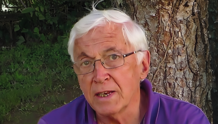
Headshot supplied by Philippe Sourdille
Over the last 50-plus years, I have observed and participated in many advancements in glaucoma surgery. But when I first took up a career analyzing the blebs of so-called filtering iridectomies, I would never have guessed that it would lead me to inventing an innovative supraciliary device. My invention is the result of a fascinating era of continuous technological and clinical advance, driven primarily by the development of microsurgery. Through magnification, for example, we gained improved visual access to the trabeculum, to Schlemm’s canal, and to the detailed, minimal modifications of aqueous outflow anatomical supports. Surgeons went from creating a hole at the corneoscleral junction to guarded filtration, trabeculectomy, non-penetrating trabecular surgery, and microinvasive glaucoma surgery. We can now take ab interno approaches aimed at direct aqueous access to Schlemm’s canal with trabecular stents or supraciliary devices to increase uveoscleral outflow. This advance came partially from John Edward Cairns’ original idea in trabeculectomy – opening Schlemm’s canal to use the existing system of outflow channels.
So, when I read about André Mermoud demonstrating different outflow supports in deep sclerectomy (1) – in particular supraciliary and suprachoroidal effusion for lowering postoperative IOP – I asked myself: “How about using a cilioscleral interposition device to ease the uveoscleral outflow?” Through scleral incisions, the device could be placed at 2 mm from the limbus, without entering the anterior chamber and without creating any subconjunctival filtration. The IOP-lowering support would be an increased supraciliary and suprachoroidal aqueous egress through the untouched ciliary band, with an additional access to intra and episcleral vessels.
In practice, of course, this initial idea presented some challenges, both technical and perceptual. Technically, it was difficult to implant the device without visual control; to control its positioning required UBM (ultrasound biomicroscopy) or OCT (optical coherence tomography). Perceptually, we faced skepticism from the broader glaucoma community about this new surgical technique.
But I was undeterred. I believed in what I was doing and enlisted support from a long-time industrial partner and friend, Olivier Benoit – a medtech and biotech entrepreneur. Together, we carefully determined the procedure’s optimal indications. After a three-year clinical follow-up, indicators revealed that our approach is effective in treating mild and moderate glaucoma. We observed a 33 percent IOP decrease and 70 percent fewer medical treatments. This three-year clinical follow-up (2) draws upon the combined results of two CIliatech studies: SAFARI I and SAFARI II with cohorts of 20 and 22 patients living with open angle glaucoma, non-controlled by medication; none had previously had glaucoma surgery. In a subsequent study (3), we enlarged the indications to angle closure glaucoma with the same surgical technique, which resulted in the same outcomes.
Our approach saw aqueous penetrate the ciliary muscle bundles through the ciliary band without a cyclodialysis cleft. This unique process needed further investigation as very few images of previous supraciliary devices are available in the literature. Successive UBM examinations were performed on all our patients during the follow-up to document the device’s position, stability, and biointegration, with an independent UBM expert analyzing all the results and confirming the device’s perfect location, stability and excellent biointegration.
We were anxious to see how glaucoma specialists reacted to this unexpected approach, and their reactions ran the gamut from skepticism to congratulations. They challenged us on the surgical technique, the peri and postoperative device position, and conjunctival involvement.
After three years, another thing I am proud of is that none of our cases have needed reintervention – but we must still consider the possibility of additional filtering procedures being needed over a patient’s lifetime, and our concept is compatible with additional procedures (if eventually needed). The concept is proven and we are now working to make the surgical technique easier, more straightforward and faster.
The last 50 years have definitely seen improvements in the benefit–risk ratio of glaucoma surgery – to the significant benefit of patients. Our research continues because, as all innovators know, innovation never ends.

Credit: Original Image from AdobeStock.com
But I was undeterred. I believed in what I was doing and enlisted support from a long-time industrial partner and friend, Olivier Benoit – a medtech and biotech entrepreneur. Together, we carefully determined the procedure’s optimal indications. After a three-year clinical follow-up, indicators revealed that our approach is effective in treating mild and moderate glaucoma. We observed a 33 percent IOP decrease and 70 percent fewer medical treatments. This three-year clinical follow-up (2) draws upon the combined results of two CIliatech studies: SAFARI I and SAFARI II with cohorts of 20 and 22 patients living with open angle glaucoma, non-controlled by medication; none had previously had glaucoma surgery. In a subsequent study (3), we enlarged the indications to angle closure glaucoma with the same surgical technique, which resulted in the same outcomes.
Our approach saw aqueous penetrate the ciliary muscle bundles through the ciliary band without a cyclodialysis cleft. This unique process needed further investigation as very few images of previous supraciliary devices are available in the literature. Successive UBM examinations were performed on all our patients during the follow-up to document the device’s position, stability, and biointegration, with an independent UBM expert analyzing all the results and confirming the device’s perfect location, stability and excellent biointegration.
We were anxious to see how glaucoma specialists reacted to this unexpected approach, and their reactions ran the gamut from skepticism to congratulations. They challenged us on the surgical technique, the peri and postoperative device position, and conjunctival involvement.
After three years, another thing I am proud of is that none of our cases have needed reintervention – but we must still consider the possibility of additional filtering procedures being needed over a patient’s lifetime, and our concept is compatible with additional procedures (if eventually needed). The concept is proven and we are now working to make the surgical technique easier, more straightforward and faster.
The last 50 years have definitely seen improvements in the benefit–risk ratio of glaucoma surgery – to the significant benefit of patients. Our research continues because, as all innovators know, innovation never ends.
References
- A Mermoud et al., “Nd:Yag goniopuncture after deep sclerectomy with collagen implant,” Ophthalmic Surg Lasers, 30, 120 (1999). PMID: 10037206.
- L. Voskanyan et al., “3 Year Outcomes of a Novel Cilio-Scleral Interposition Device (Cid Sv13): Managing IOP Without Entering the Anterior Chamber”, poster presented at the European Society for Cataract and Refractive Surgery Barcelona, September 2024.
- O Benoit, MEng, “CID and cilioscleral surgical approach clinical outcomes: 12-month follow-up results on PACG patients’ presentation delivered at the Ophthalmology futures symposium,” September 7, 2023.
