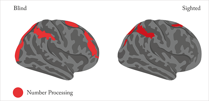
In eyecare, we often refer to “count fingers” when it comes to characterizing poor vision. But counting fingers is an example of a visual, numerical cue that helps everything from sighted, preverbal infants to non-human animals like dogs and horses learn to count. It’s known that reasoning about both approximate and exact numbers depends on a region of the brain’s cortex called the fronto-parietal network, in particular, the intraparietal sulcus (IPS). The IPS is an interesting region – it sits near the visual cortex, and is also involved in a number of aspects of vision, from saccades to depth perception. Functional MRI (fMRI) studies have suggested that IPS activity during numerical processing can be seen in children from the age of four years, and that the harder the mathematical problem, the harder the IPS works. But this begs a question: four-year olds have been counting for years before their IPS lights up on fMRI, so how much does (visual) experience – like the counting of fingers or chocolate buttons – contribute to IPS development?
To try to answer that, researchers at the Department of Psychological and Brain Sciences at Johns Hopkins University decided to use fMRI to evaluate brain activity of the whole cortices of 17 congenitally blind, and 19 blindfolded but sighted subjects (1). Both groups were subjected to spoken tests (of varying difficulty) of their mathematical and higher-level language abilities. What analysis of the fMRI data revealed was that in both blind and sighted participants, the IPS was more active during the math task than the language task (and that this activity increased parametrically with equation difficulty), suggesting that this classic fronto-parietal number network is preserved, even in the total absence of visual experience. What surprised the researchers was that blind – but not sighted subjects – also recruited a subset of early visual areas (i.e. primary visual cortex) during their symbolic mathematic calculation tests, and that the functional profile of these “visual” regions was identical to that of the IPS in blind (but not sighted) individuals. Furthermore, in the blind subjects, the regions of the visual cortex that were number-responsive – i.e. that lit up on the numerical tasks – exhibited increased functional connectivity with prefrontal and IPS regions that are known to be involved in number processing. This research reinforces previous work in blind participants which has shown that the adult visual cortex is considerably more plastic than was thought just 20 years ago (2).
References
- A Amedi et al, “Early ‘visual’ cortex activation correlates with superior verbal memory performance in the blind”, Nat Neurosci, 6, 758–766 (2003). PMID: 12808458.
