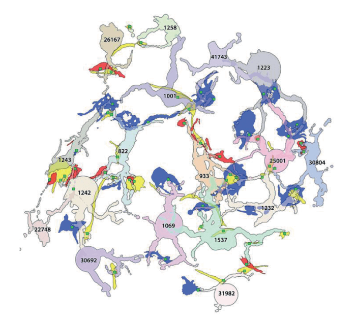
In 2011, the NIH-funded Marclab for Connectomics from the John A. Moran Eye Center at the University of Utah, Salt Lake City, USA, created a diagram of retina circuitry – the retinal connectome for vision, showing how physiology and behavior might reflect synaptic networks and their topologies. The lab then moved on to researching network changes in retinal degeneration, and has recently published a paper presenting the first ultrastructural pathoconnectome of early neurodegeneration (1).
Though the pathoconnectome has been developed based on a model of early-stage retinitis pigmentosa, the neural network changes have implications extending beyond the eye and could be helpful in researching epilepsy, Alzheimer’s, Parkinson’s and Lou Gehrig’s diseases, and even developing potential treatments. The data set used in the project is open for use by other researchers.
References
- RL Pfeiffer et al., Exp Eye Res, [Epub ahead of print] (2020). PMID: 32810483.
