Meet Leica’s Proveo 8 – a state-of-the-art ophthalmic microscope that enhances cataract surgery workflows by providing the exact image the surgeon needs at every phase of every procedure – all accessed with a simple tap of the footswitch.
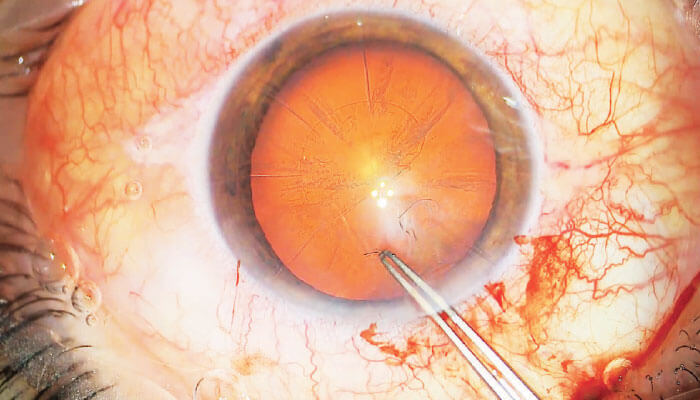
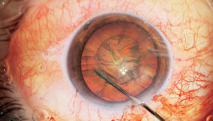
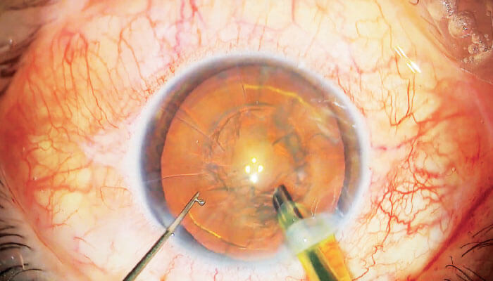
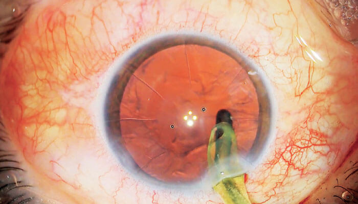
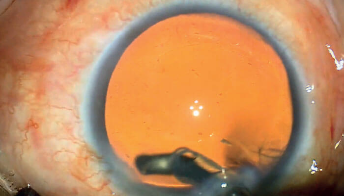
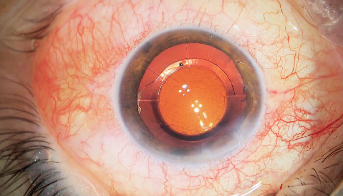
When it comes to microscopy in cataract surgery, a stable red reflex is key; by ensuring clear visibility of the lens structure, the surgeon is presented with the best view for a safe procedure. But the significant challenge with conventional systems is that red reflex illumination often diminishes when it’s needed most: during hydro dissection, phacoemulsification or cataract extraction.
Now, Leica’s new CoAx4 Illumination uses four individual beam paths to overcome this challenge, providing unmatched stable red reflex for all observers; indeed, both the surgeon and the assistant are provided with the ideal contrast for visualizing the capsule, lens, and the anterior chamber structure. Ike Ahmed, Assistant Professor and the Director of the Glaucoma and Advanced Anterior Segment Surgery fellowship at the University of Toronto, Canada, is not shy about the impact of CoAx4 Illumination: “I was captivated by Proveo’s unparalleled and consistent red reflex throughout the entire procedure.”
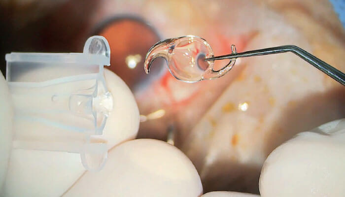
Advanced illumination is not the only trick up Proveo’s sleeve. Its unique FusionOptics boost depth of field by 40 percent – but without compromising resolution. The result? A much larger 3D visualization – in focus – which provides surgeons with clear anatomical details, right down to very small, delicate structures.
The Proveo has also been optimized for use in busy practices. It includes separate user settings for up to 30 user profiles, a feature that not only recognizes the fact that each surgeon has unique ergonomic preferences for maximum comfort – but it also saves valuable set-up time.
Another time saving and workflow enhancing feature is the Combination Mode, which allows surgeons to pre-define and program specific levels of light, focus and magnification for up to five procedure phases (for example, capsulorhexis, phacoemulsification, irrigation/aspiration, posterior capsule polishing, IOL positioning in cataract surgery).
And how are the programmed settings for the next phase activated? “With the Proveo 8, I customize the light settings by a simple click on the foot switch,” explains Devesh Varma, Assistant Professor in the Department of Ophthalmology and Vision Sciences at the University of Toronto.
And there’s no need for users to reset all the major functions of the microscope (zoom, focus, light and XY) to zero – the Proveo 8 performs the task automatically, resulting in further time savings.
As a busy cataract surgeon, you want to make better-informed decisions – fast. By simplifying access to exactly the right image at exactly the right time, the Proveo 8, with its host of advanced features, helps avoid interruptions and streamlines your workflow.
Raise the bar now.
