The SPECTRALIS® High Magnification Module (HMM) is designed to visualize significantly more microstructural detail of the ocular fundus than is possible with a standard SPECTRALIS lens. Far from being a simple digital zoom, the HMM capitalizes on the exceptional SPECTRALIS technology by sampling with particularly high density.
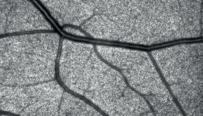
How? The non-invasive HMM makes optimal use of SPECTRALIS’ confocal scanning laser ophthalmoscopy (cSLO) technology by combining it with the selectivity of laser light to resolve retinal microstructures with a level of detail typically not available in a traditional clinical setting. Such detail could provide novel insights into the pathogenesis and progression of retinal diseases, thus helping ophthalmologists refine surgical and treatment regimens.
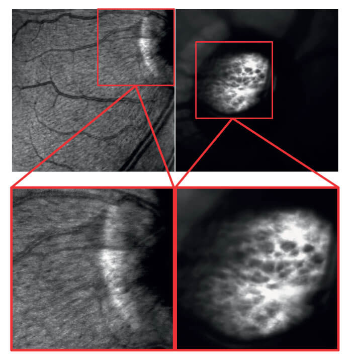
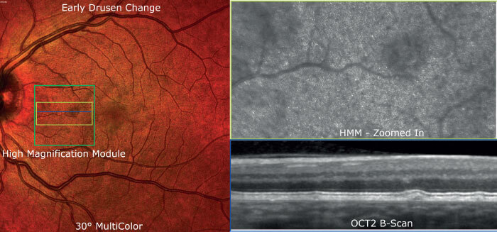
The SPECTRALIS multimodal imaging platform offers the flexibility to identify a region of interest with the standard 30° field of view, the widefield 55° or the ultra-widefield 102° – and then magnify that region using the HMM to obtain high-resolution 8° x 8° infrared reflectance images. The magnification generates much denser scans of the region, presenting highly detailed information of ocular microstructures. The field of view of the HMM allows for closer investigation of the areas of interest while still being large enough to aid navigation and orientation.
Case study: Clinical applications in a multimodal context
Early adopters of this new technology are currently combining the HMM with more established imaging modalities to examine patients with ellipsoid zone disruption and outer retinal changes, early drusen and drusen-like deposits in the context of AMD, and pachychoroid disease, as well as for patients with nerve fiber bundle defects and for the assessment of retinal vessel walls. The HMM images reveal details of retinal microstructures that could remain unseen with most imaging modalities.
Giovanni Staurenghi, Professor of Ophthalmology at the University of Milan, Italy, said, “It’s promising to be able to obtain infrared fundus images with such a degree of magnification using our regular SPECTRALIS device. It seems like we are now seeing down to the photoreceptor level. We have just started using the High Magnification Module as part of our multimodal imaging approach and we are looking forward to exploring its full clinical value.”