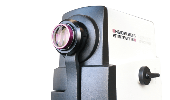
The Heidelberg Engineering SPECTRALIS® is more than just a multimodal imaging platform – it’s an invaluable technology in the fight against AMD and other retinal diseases. Optimized for the posterior segment of the eye, its core technologies guarantee exceptional image quality, consistently delivering the information needed for accurate patient assessment alongside the precision required for long-term follow-up examinations.
The SPECTRALIS draws on cutting-edge confocal scanning laser ophthalmoscopy (cSLO) technology to provide high-contrast, high-quality images – even in the most challenging patients. Combining the selectivity of laser light with the resolution of confocal scanning, the SPECTRALIS guarantees a level of image detail and clarity that cannot be achieved with fundus photography alone. It also offers live fundus assessment with simultaneously captured OCT images, enabling users to pinpoint pathology with minimal light scatter thanks to the unique properties of cSLO imaging. It can even be used in patients with cataracts.
Freeze time
TruTrack Active Eye Tracking is a patented technology that uses a secondary laser beam to actively track the eye during OCT scanning to avoid motion artifacts. TruTrack effectively “freezes” the retina, allowing the operator to capture precise OCT images even when the patient blinks or moves. The benefits of this are twofold: the patient is comfortable and the doctor is confident that the scan data are accurate. Beyond those core benefits, TruTrack Active Eye Tracking allows precise, follow-up scanning and excellent image quality throughout the volume scan, even in cases where there are relatively small changes over time.
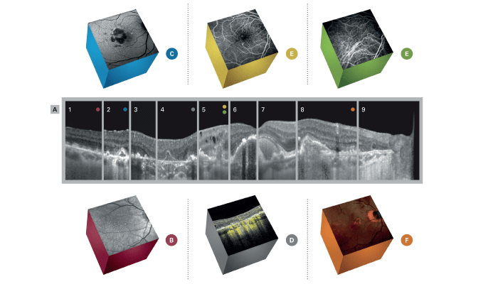
A. Structural OCT
1. Subretinal drusenoid deposits
2. Calcified drusen
3. Descent of the external limiting membrane
4. Persistent basal laminar deposits in the atrophy bed
5. Type 3 macular neovascularization
6. Drusenoid pigment epithelial detachment
7. Acquired vitelliform lesion
8. Type 1 macular neovascularization
9. Outer retinal tubulation
B. Near-infrared reflectance with OCT imaging (1) Near-infrared reflectance enables a comprehensive overview of the fundus and serves as a reference image when combined with OCT. It shows useful information on pathologic changes, as seen in this case of subretinal drusenoid deposits, observed as hyporeflective dots all over the macula.
C. BluePeak autofluorescence (2) Blue fundus autofluorescence with the BluePeak Module gives information about the status of the retinal pigment epithelium (RPE), showing areas of complete atrophy of the outer retina and the RPE, as seen in this case of calcified drusen.
D. OCT angiography (4) Images captured with the OCT Angiography Module show flow signal over the structural information of the OCT scan, supporting the differential diagnosis of hyporeflective structures within the outer retina. OCTA images can help identify neovascularization, as in this case of persistent basal laminar deposits in an area of RPE and outer retinal atrophy.
E. Fluorescein & indocyanine green angiography (5) In combination with structural OCT and OCTA, dye-based angiography provides a detailed analysis of the vascular architecture of the retina and the choroid, as in this case of type 3 macular neovascularization or retinal angiomatous proliferation.
F. MultiColor (8) MultiColor reflectance uses three lasers with different wavelengths to create a combined image that provides information about different levels of the retina and choroid; longer wavelengths enable higher penetration depth. This case shows a deep lesion with orange borders, corresponding to a type 1 macular neovascularization.
Christine A. Curcio, Director of the AMD Histopathology Lab of the Department of Ophthalmology and Visual Sciences, University of Alabama at Birmingham, USA
“AMD could be beat if ophthalmologists could use everything that is in the OCT B-scans. The secret to accurate diagnosis of manifold retinal diseases often lies in OCT images. This, of course, is highly influenced by the quality of the obtained images and requires a high level of clarity and contrast, as well as the ability to repeatedly capture the exact same spot where the disease is estimated.”

Rosa Dolz-Marco, Retina Specialist, Oftalvist Clinic, Valencia, Spain
“The wide range of imaging modalities combined within the SPECTRALIS gives me certainty and confidence when diagnosing a broad range of retinal diseases and abnormalities. The modalities complement each other facilitating precise examinations and follow-ups. In a busy clinic, it is also very convenient that the patient doesn’t need to move between too many devices and feels comfortable during the examination.”

References
- L Chen et al., Retina. 40, 618 (2020). PMID: 31599795.
- ACS Tan, Sci Transl Med, 7, 10, 466 (2018). PMID: 30404862.
- R Dolz-Marco, Am J Ophthalmol, 193, 166 (2018). PMID: 29981740.
- ACS Tan, Invest Ophthalmol Vis Sci, 1, 58, 2349 (2017). PMID: 28437524.
- M Li, Ophthalmology, 125, 276 (2018). PMID: 28964579.
- C Balaratnasingam, Invest Ophthalmol Vis Sci, 1, 57, 5479 (2016). PMID: 27760262.
- KC Chen, Am J Ophthalmol, 164, 89 (2016). PMID: 26868959.
- C Curcio, Am J Ophthalmol, 160, 1024 (2015). PMID: 26255578.
- R Dolz-Marco et al, Ophthalmology, 124, 1353 (2017). PMID: 28456420.
