In just 30 years, Optos technology has evolved from a hand-drawn diagram on a paper napkin to a breakthrough innovation that is transforming the fight against vision loss: optomap® ultra-widefield retinal imaging. This technology is integral to Silverstone, the only retinal imaging device to integrate ultra-widefield capabilities with swept-source OCT, and which can produce a 200° single-capture retinal image of unrivalled clarity in <0.5 seconds.
How and why are these tools changing clinical practice? We listened to Paulo Stanga, retina surgeon at the Retina Clinic London and London Vision Clinic, UK, Tunde Peto, Professor of Clinical Ophthalmology at Queen’s University Belfast, UK, and SriniVas Sadda, Director of Artificial Intelligence & Imaging Research, Doheny Eye Institute, and Professor of Ophthalmology at UCLA, California, USA, as they gave their updates at the recent Euretina congress.
Integrating ultra-widefield imaging with navigated SS-OCT in the retina clinic: real life benefits
Paulo Stanga
The Optos Silverstone combination of ultra-widefield (UWF) imaging with navigated SS-OCT is unique – but what are the real-world benefits? Key advantages, says Stanga, include the ability to generate high resolution images from not only posterior but also mid and peripheral retina, generating a cross sectional OCT image of 80 degrees (23 mm) in length. The regular OCT is 23 mm: much larger than what most SD-OCT devices are capable of, and competitive with other SS-OCT. SS-OCT capabilities, he adds, permit assessment of the deeper ocular regions – essential for assessing myope retinas. Furthermore, Silverstone provides unparalleled visualisation of the vitreoretinal interface: “This allows optimal PVD assessment, which is essential now that floaters are treated with laser and limited vitrectomy.” Silverstone thus combines SS-OCT advantages – for example, in visualizing sub-retinal fluid and guiding management of retinal detachments, peripheral retinal tears, and 360-degree retinoplexy – with the complementary benefits of UWF imaging. The clinical utility of Silverstone is therefore clear (see Box 1).
In brief, says Stanga, peripheral OCT scanning is essential in clinics where indirect ophthalmoscopy with 360-degree scleral indentation is unavailable, while UWF imaging is increasingly important in disease diagnosis and management. Combining the two imaging approaches allows assessment of both peripheral retina and vitreoretinal interface, and so aids diagnosis of vitreoretinal pathologies. He concludes: “All my patients undergo UWF imaging – I can’t imagine my retina clinic without it.”
Leveraging Optos UWF imaging technology for AI-based DR grading
Tunde Peto
An optomap-based system has now joined the exclusive club of approved AI-based ophthalmological diagnostics – the fruit of a massive collaboration (see Sidebar 2) between several parties, including Google and numerous grading centers. This was an enormous undertaking, notes Peto: “Double-grading ultra-widefield images from 67,000 patients generates a lot of data!” Furthermore, she adds, the grading system examined both the standard seven fields and also extensions of fields 3-7 to ensure capture of predominantly peripheral lesions (PPLs): “This was necessary to optimize data for algorithm training – but resulted in each image being associated with 179 individual fields!”
The rigor implied by the above was evident throughout the project. For example, since accuracy of AI algorithms can be affected by variations in background pigmentation, images were chosen to ensure coverage of different ethnic backgrounds. Furthermore, the project incorporated internal controls to assess intra-grader consistency: 33 percent of graders who graded >100 images were sampled, and up to 5 percent of their graded images were randomly selected and returned to each grader’s work-list in a masked fashion. Re-grades had over 80 percent agreement with original grades, demonstrating high intra-grader consistency.
This enormous effort resulted in an AI-led, UWF-based grading tool with 96.1 percent sensitivity and 93.4 percent specificity. The system is already CE-marked and commercially available in the in the UK and EU, and should soon be available in the US. Peto concludes: “Our algorithm will continually learn from all the images the global ophthalmology community give it to analyze – it will just get better and better!”
Peripheral diabetic retinopathy lesions as identified by UWF imaging – changing clinical practice?
SriniVas Sadda
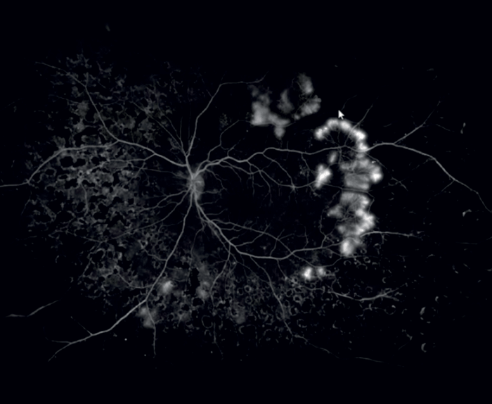
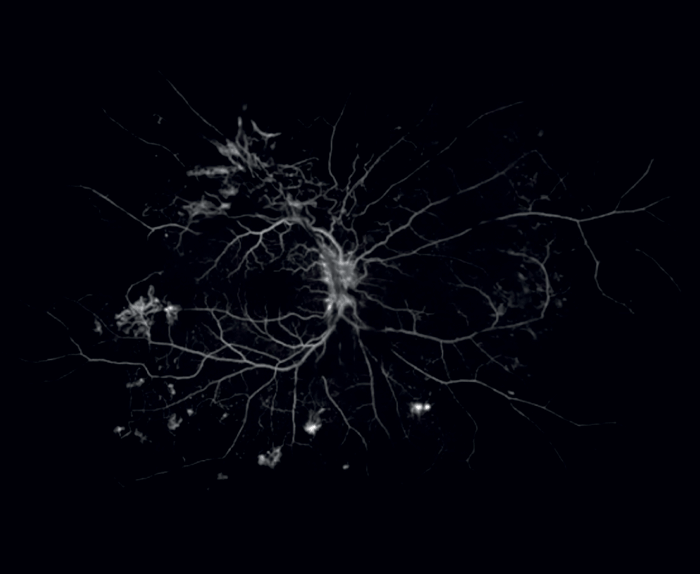
A key question in the management of diabetic retinopathy (DR) is: can lesions outside the seven standard fields – predominantly peripheral lesions (PPLs) – predict disease progression? As Sadda says, “This would have far-reaching implications for patient management and follow-up, and could provide new insights into DR.” Fortunately, the recent DRCR Retina Network Protocol AA study has provided some answers.
Firstly, protocol AA demonstrates that patients can have relatively mild disease in the posterior pole but extensive pathology in the periphery. Secondly, and most importantly, the study shows that patients with PPLs identified by UWF-FA are at 1.7- fold higher risk of DR progression, over four years, than patients in whom UWF-FA does not reveal PPLs. W ith UWF-color assessments, this relationship holds only in moderately severe and severe and PDR patients. Therefore, in eyes with non-proliferative disease, UWF-FA evaluation can signal progression risks that are not detectable via UWF-color evaluation, assessment of the seven standard fields, OCT or OCTA. This association of PPLs with progression risk has important implications for disease management.
Conclusions
The long-awaited combination of UWF-FA and SS-OCT, manifest in the Optos Silverstone, is transforming ophthalmological practice. Complementary information provided by these technologies – notably, the finding that PPLs identified by UWF-FA predict risk of DR progression – will guide disease management. The new AI-based DR grading algorithm – trained on UWF images – will bring a new level of sensitivity and specificity to the clinic. In particular, the real-time feedback provided by Optos AI for DR enables physicians to immediately determine whether a patient should be referred for further DR evaluation and management. Finally, we should not overlook the convenience of Optos imaging, which provides as much information in a single UWF image as can be collected from seven standard fields. Fewer images mean faster throughput and greater patient convenience – key advantages in modern ophthalmology. In brief, ultra-widefield is a powerful diagnostic tool; the high quality, clinically meaningful images it generates are changing DR clinical management to the great benefit of physicians and patients.
