Disclaimer: The High-Res OCT is an investigational device not available for sale. For more information, contact retina@HeidelbergEngineering.com.
The Heidelberg Engineering SPECTRALIS® is an internationally renowned OCT platform, which combines confocal Scanning Laser Ophthalmoscopy (cSLO) with OCT, OCT angiography, dye-based angiography, and MultiColor imaging. One of the latest innovations – the High-Res OCT – is an investigational device with a light-source capable of providing an axial resolution of up to 3 µm that could bring researchers new insights into the retinal structure and vasculature. This investigational device is based on the existing SPECTRALIS multimodal imaging platform, with one major difference; the High-Res OCT generates greater axial resolution and allows clinicians to see more detail.
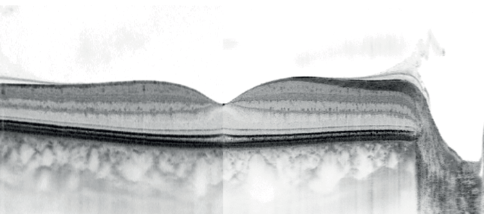
The optical axial resolution of an OCT system depends on two light source properties: the bandwidth and the central wavelength. To achieve a greater optical axial resolution with a longer central wavelength (for example, 1060 nm), a larger bandwidth will be required when compared with a near-infrared (NIR) or visible spectrum. The High-Res OCT works with a central wavelength of 840 nm and an increased bandwidth, making it possible to improve the optical axial resolution in tissue from 7 to 3 µm.
Increasing the bandwidth of the light source instantly improves the optical axial resolution and, in turn, reproduces the finest details of the eye. SD-OCT is the only current technology capable of achieving a significantly higher axial resolution and presently, there are no swept-source light sources with higher bandwidth available to allow the development of an SS-OCT device with increased axial resolution.
Over the last 30 years, OCT has become a powerful diagnostic tool by enabling non-invasive visualization of the posterior and anterior segments of the eye. The first OCT devices employed time-domain OCT (TD-OCT), which were shortly followed by spectral-domain OCT (SD-OCT) and swept-source OCT (SS-OCT) devices, which provided significantly higher speed, higher resolution, and contrast. SD-OCT and SS-OCT are both technologies that operate in the frequency domain.
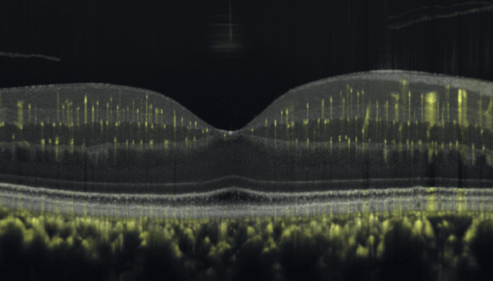
SS-OCT coupled with a longer wavelength delivers the advantage of deeper optical depth penetration, while SD-OCT, used with a shorter wavelength, can provide higher contrast within the retina and superior optical axial resolution. This combination is particularly suited for visualization of the inner and outer retina to Bruch’s membrane, resulting in stunning images of great clarity and contrast.
To learn more about this topic, take a look at the paper “State-of-the-art commercial Spectral-Domain and SweptSource OCT technologies and their clinical applications in ophthalmology” at www.spectralis.info.
The improved axial resolution of the High-Res OCT of 3 µm results in clearer and more detailed images resolving even minuscule vessels in the retinal layers and the choriocapillaris. The High-Res OCT with its broader OCT light spectrum has the potential to provide clinicians with new enlightening insights.
