Conexiant
Login
  • Corneal Physician
  • Glaucoma Physician
  • New Retinal Physician
  • Ophthalmology Management
  • Ophthalmic Professional
  • Presbyopia Physician
  • Retinal Physician
The Ophthalmologist
  • Explore

    Explore

    • Latest
    • Insights
    • Case Studies
    • Opinion & Personal Narratives
    • Research & Innovations
    • Product Profiles

    Featured Topics

    • Anterior Segment
    • Glaucoma
    • Retina

    Issues

    • Latest Issue
    • Archive
  • Subspecialties
    • Cataract
    • Cornea
    • Glaucoma
    • Neuro-ophthalmology
    • Oculoplastics
    • Pediatric
    • Retina
  • Business

    Business & Profession

    • Professional Development
    • Business and Entrepreneurship
    • Practice Management
    • Health Economics & Policy
  • Training & Education

    Career Development

    • Professional Development
    • Career Pathways

    Events

    • Webinars
    • Live Events
  • Events
    • Live Events
    • Webinars
  • Community

    People & Profiles

    • Power List
    • Voices in the Community
    • Authors & Contributors
  • Multimedia
    • Video
    • Podcasts
Subscribe
Subscribe

False

Advertisement
The Ophthalmologist / Issues / 2021 / Oct / Image of the Month: Intralenticular Foreign Body

Image of the Month: Intralenticular Foreign Body

Intralenticular Foreign Body

By Geoffrey Potjewyd, Aleksandra Jones 10/15/2021 1 min read

Share

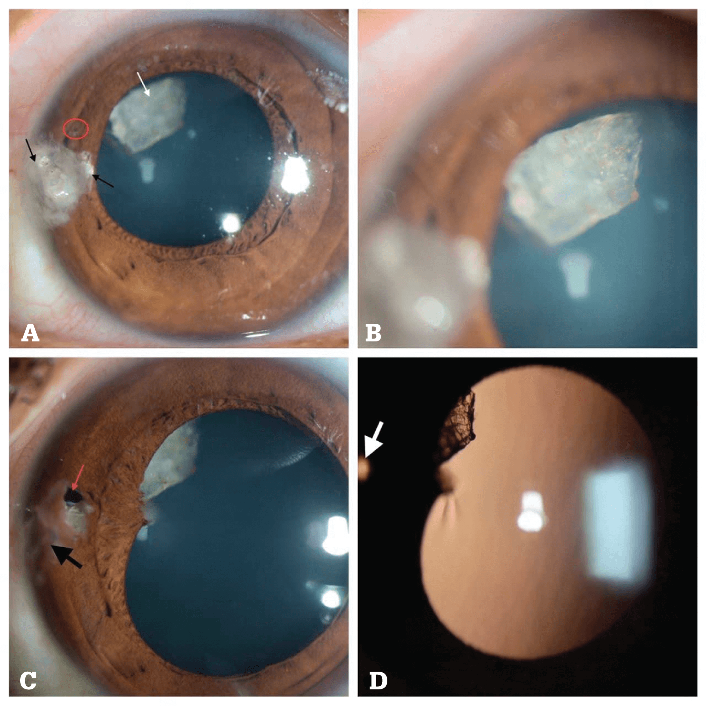
(A) Preoperative image of sealed corneal laceration with foreign body pieces and mucus embedded within the wound (thin black arrows); intralenticular foreign body (ILFB) seen out of focus (thin white arrow); though difficult to appreciate, a small iris hole is noted corresponding to the entry wound (within red circle).
(B) Magnified view showing the refractile nature of the ILFB.
(C) One month post-op, the healed wound is seen after suture removal (thick black arrow); iris defect is appreciated now (red arrow) with atrophy of iris tissue around it; the inert ILFB is partially obscured by focal posterior synechia. The remaining lens is clear, and there is no intraocular inflammation.
(D) Retroillumination view showing the ILFB partially obscured by posterior synechia and transillumination at the site of iris defect (thick white arrow).

Credit: Raksheeth Nathan Rajagopal and Muralidhar Ramappa, LV Prasad Eye Institute, Hyderabad, India.

References

  1. RN Rajagopal, M Ramappa, BMJ Case Rep, 14, e244875. PMID: 34479900.

About the Author(s)

Geoffrey Potjewyd

The lion’s share of my PhD was spent in the lab, and though I mostly enjoyed it (mostly), what I particularly liked was the opportunity to learn about the latest breakthroughs in research. Communicating science to a wider audience allows me to scratch that itch without working all week only to find my stem cell culture has given up the ghost on the Friday (I’m not bitter). Fortunately for me, it turns out writing is actually fun – so by working for Texere I get to do it every day, whilst still being an active member of the clinical and research community.

More Articles by Geoffrey Potjewyd

Aleksandra Jones

Having edited several technical publications over the last decade, I crossed paths with quite a few of Texere's current team members, and I only ever heard them sing the company's praises. When an opportunity arose to join Texere, I jumped at the chance! With a background in literature, I love the company's ethos of producing genuinely engaging content, and the fact that it is so well received by our readers makes it even more rewarding.

More Articles by Aleksandra Jones

Related Content

Newsletters

Receive the latest Ophthalmology news, personalities, education, and career development – weekly to your inbox.

Newsletter Signup Image

False

Advertisement

False

Advertisement

Explore More in Ophthalmology

Dive deeper into the world of Ophthalmology. Explore the latest articles, case studies, expert insights, and groundbreaking research.

False

Advertisement
The Ophthalmologist
Subscribe

About

  • About Us
  • Work at Conexiant Europe
  • Terms and Conditions
  • Privacy Policy
  • Advertise With Us
  • Contact Us

Copyright © 2025 Texere Publishing Limited (trading as Conexiant), with registered number 08113419 whose registered office is at Booths No. 1, Booths Park, Chelford Road, Knutsford, England, WA16 8GS.

Disclaimer

The Ophthalmologist website is intended solely for the eyes of healthcare professionals. Please confirm below: