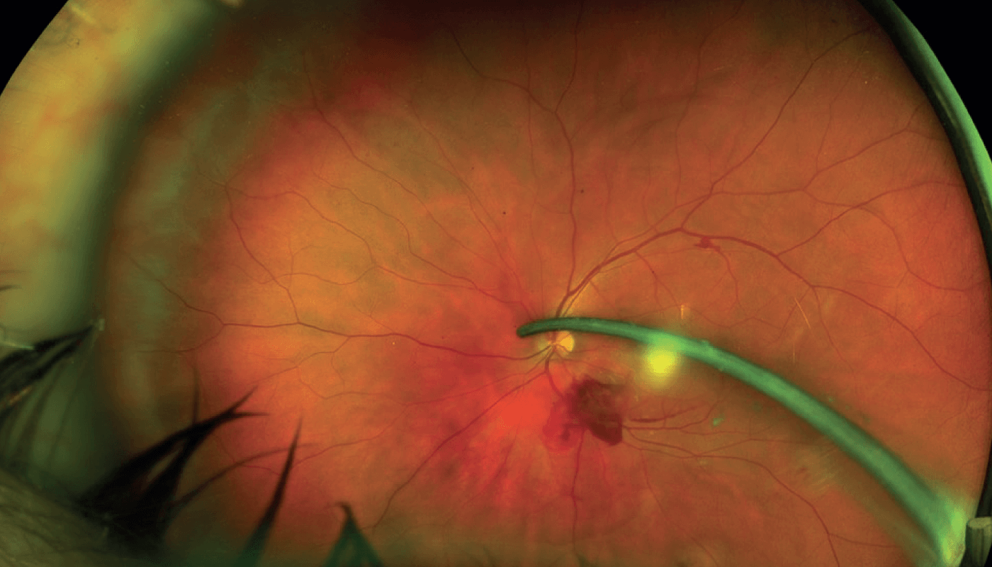
Image of the Month: A Foreign Body
A metallic intraocular foreign body very close to the optic nerve
This month’s image shows a metallic intraocular foreign body very close to the optic nerve. It was removed safely, despite initial immediate postop vitreous inflammation, and the patient recovered completely within three weeks, with unaided visual acuity of 20/20.
Credit: Costas H. Karabatsas, Assistant Professor of Ophthalmology at the Department of Biomedical Sciences of the University of West Attica, Psachna, Greece.
Having edited several technical publications over the last decade, I crossed paths with quite a few of Texere's current team members, and I only ever heard them sing the company's praises. When an opportunity arose to join Texere, I jumped at the chance! With a background in literature, I love the company's ethos of producing genuinely engaging content, and the fact that it is so well received by our readers makes it even more rewarding.