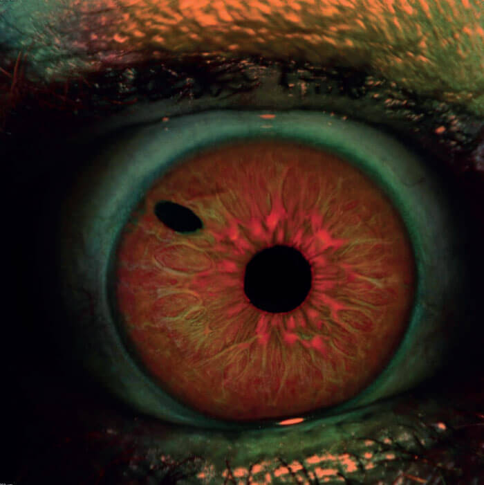
The MultiColor scanning laser doesn’t take traditional fundus images, but it can show structures with pathologies not visible when using ophthalmoscopy and fundus photography. Three individual laser wavelengths: blue, green, and infrared, are simultaneously captured to create the image. This iris image was taken a couple of weeks after the patient’s YAG laser peripheral iridotomy.
Credit: Stephanie Moolman, Ophthalmic Photographer/Technician, South Africa
Read the full article now
Log in or register to read this article in full and gain access to The Ophthalmologist’s entire content archive. It’s FREE and always will be!
Login
Or register now - it’s free and always will be!
You will benefit from:
- Unlimited access to ALL articles
- News, interviews & opinions from leading industry experts
- Receive print (and PDF) copies of The Ophthalmologist magazine
Or Login via Social Media
By clicking on any of the above social media links, you are agreeing to our Privacy Notice.
About the Author
